

MiWord of the Day Is… Haptoglobin!


Just got back from the RSNA! Wow what a big conference – 56,000 people this year. McCormick place in Chicago, Illinois (where the conference is held) feels like an airport it is so big. 


Love Chicago. Great city. 


Of course, I had the pleasure of attending a bunch of great presentations and today I will introduce you to one of them. Tina Binesh Marvasti (say that 7 times fast!) presented on the topic of Haptoglobin. No, not Hobgoblin (not sure who that is? See here) or his infamous green predecessor (see here). 


So, what is Haptoglobin you ask? It is a serum protein that binds free hemoglobin – resulting from the breakdown of red blood cells – and functions to prevent loss of iron (contained in the heme group) through the kidneys and to protect tissues from the highly reactive heme groups. Essentially a housekeeping protein that helps to recycle hemoglobin as part of the red blood cell life cycle. Now what if your ability to clean-up free hemoglobin was impaired? Well, quite simply you would be putting at risk those sensitive tissues that come into contact with free hemoglobin. 
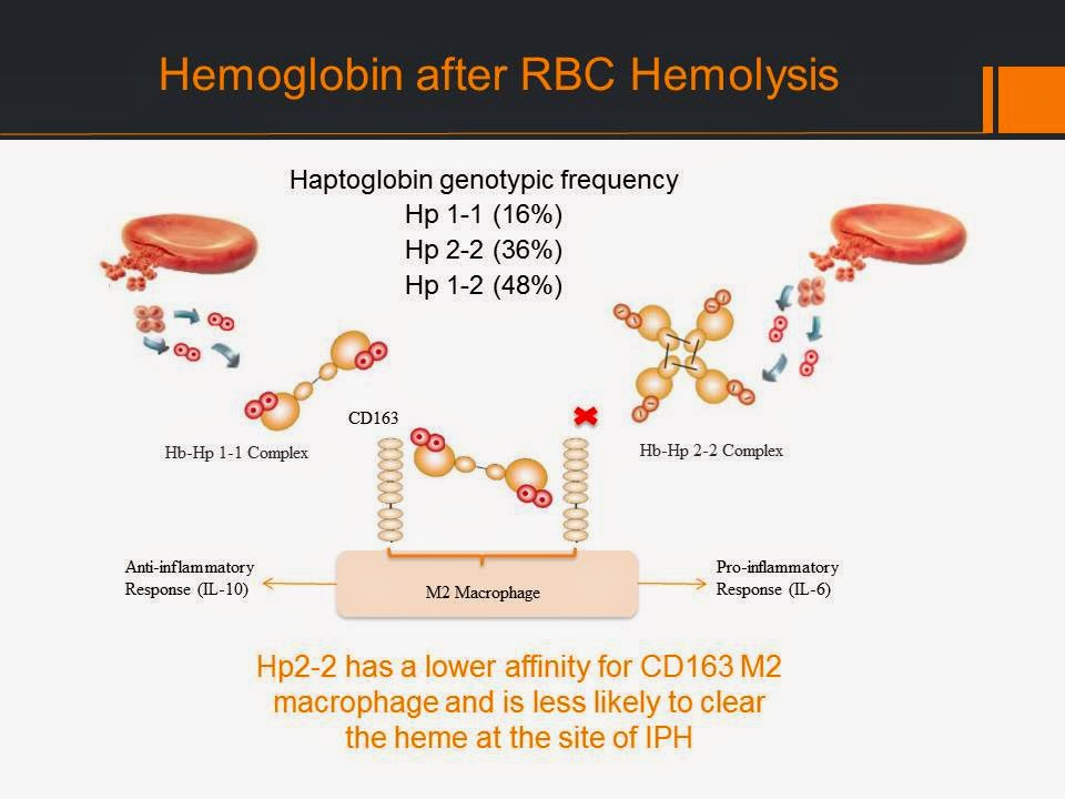


One important example of this is vessel walls affected by atheroma (AKA plaque). Sometimes these atheroma can bleed (called intraplaque hemorrhage or IPH) which worsens the whole situation. Typically, your body responds by sending the clean-up crew including the Hobgoblin (or haptoglobin, I always get these two confused). 


When people have the recessive genotype (Hp 2-2) of the Hp gene they produce less haptoglobin and therefore are at increased risk of damage from free hemoglobin (or more specifically the heme groups).


Tina and friends hypothesized the following: 


And she found that having the recessive Hp2-2 genotype was associated with a higher prevalence of IPH in a group of  80 patients (average age of 73 yrs). She also found that the IPH volume of Hp2-2 patients worsened over time.


So what is the take home? The Haptoglobin genotype is associated with IPH which is a biomarker of high risk vascular disease and could identify populations at higher risk of developing cardiovascular events.


Now for the fun part (see the rules here), using Haptoglobin in a sentence by the end of the day:

Serious: Hey Bob, did you know that a recessive haptoglobin genetype may contribute to an increased risk of cerebrovascular disease?

Less serious: My GP suggested that based on my recessive hobgoblin genotype I should consider a healthier lifestyle. Funny, I always figured Doc Ock to be the one to watch for…

OK, watch the Spider-man 2 trailer to decompress and I’ll see you in the blogosphere…


Pascal Tyrrell

MiWord of the Day Is… CAT scan!

A what scan? I am actually a cat guy myself. Not to say I don’t love dogs but if I had to make a choice…


I just finished reading a fantastic book by David Dosa entitled “Making Rounds With Oscar”. The premise of the book is a story about an extraordinary cat but the subject matter is very serious –  dementia and end-of-life care in the elderly. Have a gander.


So what the heck is a cat scan and what does it have to do with medical imaging? 


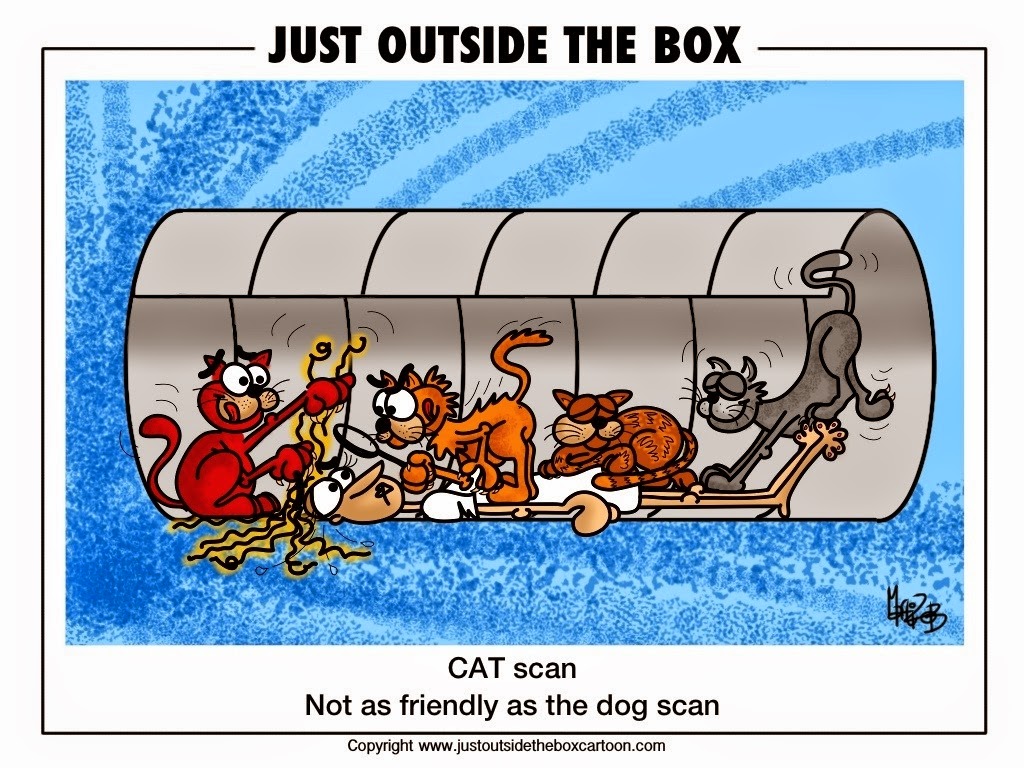
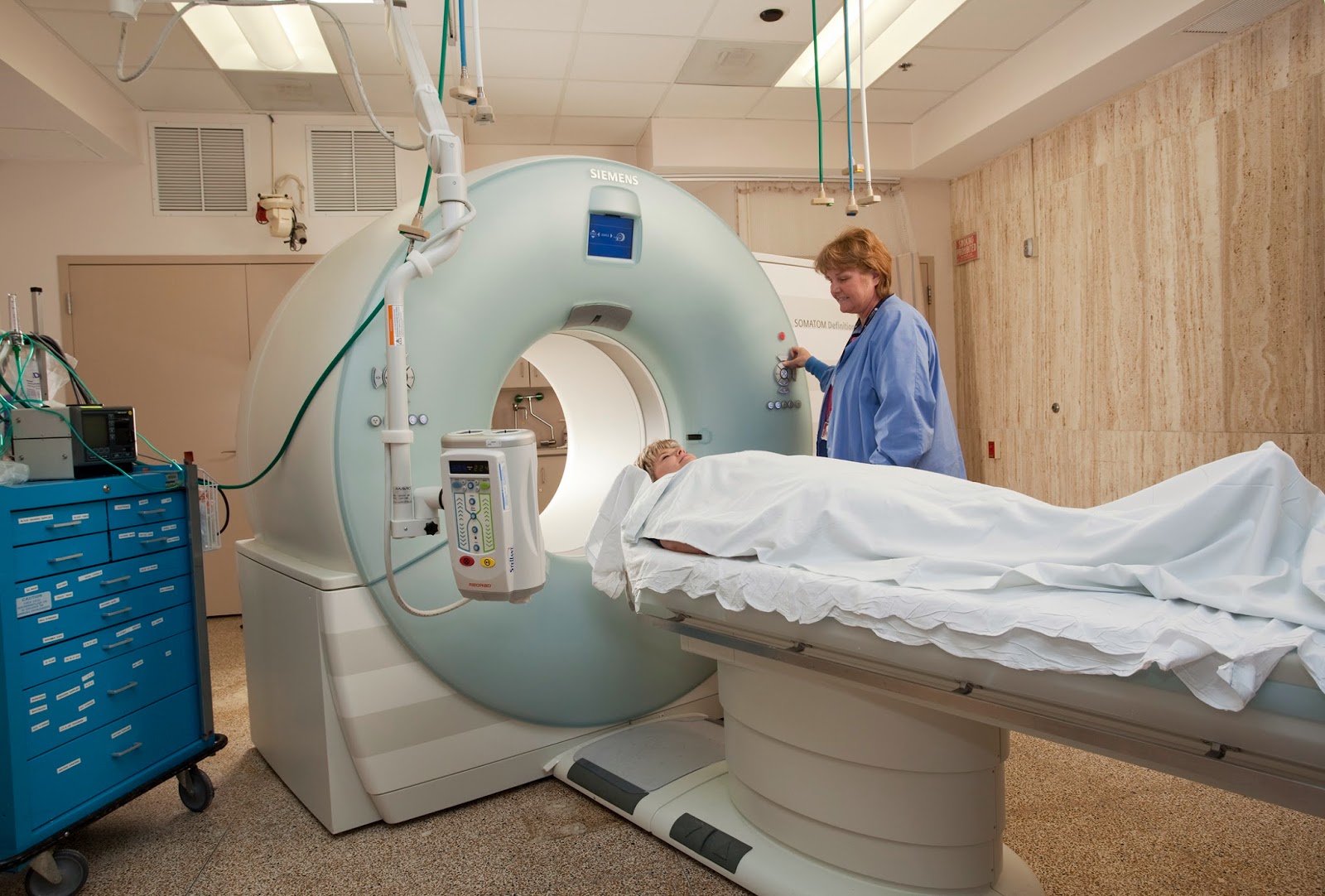
CT scans – also referred to as computerized axial tomography (CAT) – are special X-ray tests that produce cross-sectional images of the body using X-rays and a computer. CT was developed independently by a British engineer named Sir Godfrey Hounsfield and Dr. Alan Cormack and were jointly awarded the Nobel Prize in 1979. Yes, more Nobel prize winners…


In a nutshell, x-ray computed tomography:


– uses data from several X-ray images of structures inside the body and converts them into 3D pictures – especially useful for soft tissues.


– emits a series of narrow beams through the human body, producing more detail than standard single beam X-rays.


– is able to distinguish tissues inside a solid organ. A CT scan is able to illustrate organ tear and organ injury quickly and so is often used for accident victims.


– is analyzed by radiologists.


Unfortunately, unlike MRI scans, a CT scan uses X-rays and therefore are a source of ionizing radiation.


Now for the fun part (see the rules here), using CAT Scan in a sentence by the end of the day:

Serious: Hey Bob, did you know that the recorded image of a CAT Scan is called a tomogram? 

Less serious: My GP suggested that howling at the moon at night is not normal behavior and he wants to send me for a CAT scan. What? No way, I’m allergic to cats…

OK, listen to Cat Stevens to decompress and I’ll see you in the blogosphere…

 
Pascal Tyrrell

MiWord of the day is… Atom!

MiWord, a post on Sunday?!!! Well, I have been very busy lately and fell behind on my blog so I am now playing a little catch-up…


I was waiting in Logan airport for my flight back from a presentation in Boston – what unbelievably crazy traffic in that city – and I was texting my kids with my laptop open and my tablet next to me on the seat when I thought: I feel a little like Jimmy Neutron! I enjoyed watching that show with my kids. Lots of fun. Anyway, that idea of crazy science and the internal structure of the atom as displayed on Jimmy’s t-shirt may be the premise for a great kids show but it also led to the development of MRI. What?!!! You say. 


MRI is an imaging technique. Maybe so, but it is particular in that it does not use any classic photographic equipment (film or lenses) or use x-rays as Roentgen did. It simply numerically measures how hydrogen nuclei absorb and release energy in response to particular frequencies. Need a refresher on the structure of an atom? See this post.


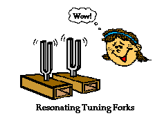
Don’t get it? OK, how about you think of this process as a crazy huge tuning fork. If you were to flick a tuning fork of a certain frequency (pitch) other tuning forks of the same frequency close by will pick up energy from the humming tuning fork and emit a sound in turn. Cool.


The nucleus of an atom can absorb energy and then relax by emitting energy in a similar way. Different atoms (or the same atom in different environments) will have different relaxation rates allowing for the identification of the composition of molecules. Ya, maybe a little complicated.


MRI measures how hydrogen nuclei absorb and release energy. Dependent on the location and the environment of the hydrogen atoms the MRI process is able to provide knowledge about the placement of hydrogen atoms in the body and in turn knowledge about the anatomy.


Now for the fun part (see the rules here), using Atom in a sentence by the end of the day:

Serious: Hey Bob, did you know that the atom is the smallest unit that defines the chemical elements and their isotopes?

Less serious: I thought that splitting atoms would produce a large explosion but when I tried using my mom’s perfume “atomizer” it just produced a fine spray and nice smell…


Ok that was a little intense for a Sunday. Watch and listen to Symphony of Science (very cool BBC production) to decompress and I’ll see you in the blogosphere…



Pascal Tyrrell

MiWord of the Day Is… Cinemaradiology!

Yes, it is Halloween today and my kids could barely contain themselves getting ready for school. I suspect today will not be very productive as they count down the minutes before heading out to terrorize my neighbors.


Anyway, how about this for a scary thought: cinemaradiology! In the late 1800’s John MacIntyre at the Gasgow Royal Infirmary experimented with producing X-ray motion pictures. What!!!? He tried exposing film by passing it between the screen of the fluoroscope and the x-ray tube and by simply filming the fluoroscopic screen. This latter method was very difficult because, as all of you budding radiologists know, the images viewed on the fluoroscope screen were dim and of poor resolution at the time.


For years researchers worked on perfecting cinemaradiology. However, during those early years of discovery they lost interest when they realized that sharper images were possible when BOTH patients and investigators were exposed together AND that excessive radiation was a bad thing – duh! 


It would only be many many years later that fluoroscope screen technology would be improved to allow for brighter and higher resolution images (and without frying the patient and everyone around!). 

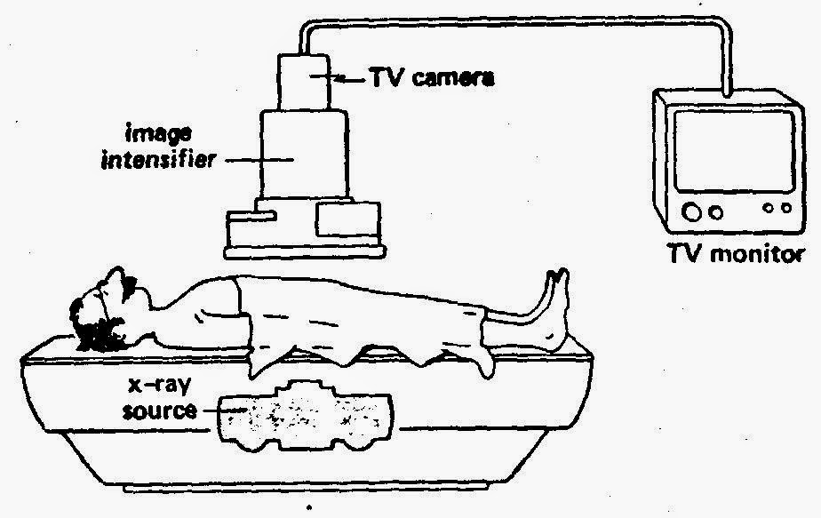
Fluoroscopy is a study of moving body structures – similar to an x-ray “movie.”  A continuous x-ray beam is passed through the body part being examined, and is transmitted to a TV-like monitor so that the body part and its motion can be seen in detail. As an imaging tool, Fluoroscopy is used in many types of examinations and procedures.

To my knowledge, no actors from the cinemaradiology era ever became successful stars in Hollywood…


No need to use the MiWord of the day in a sentence today (see rules here) as I realize you are busy getting ready for Halloween and need a break!


Decompress listening to the classic song Thriller by the King of Pop Michael Jackson and I’ll see you in the blogosphere…


Pascal Tyrrell


What Cluster Are You From?

This week and I had the pleasure of presenting to the Division of Rheumatology Research Rounds – University of Toronto. They were a fantastic audience who asked questions and appeared to be very engaged. Shout out to the Rheumatology gang!


So, I was asked to talk about a statistical methodology called Cluster Analysis. I thought I would start a short series on the topic for you guys. Don’t worry I will keep the stats to a minimum as I always do!


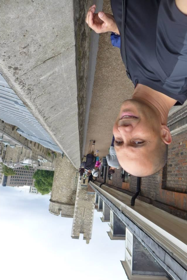
Complex information can always be best recognized as patterns. The first picture below on the left certainly helps you realize that it is not a simple task to know someone at a glance.


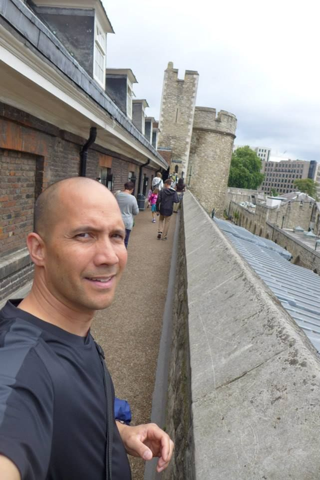
Now, I guess it doesn’t help that many of you have never met me either! However, you can appreciate that things get a little easier when the same portrait is presented in the usual manner – upright! 


This is an interesting example where the information is identical, however, our ability to intuitively recognize a pattern (me!) appears to be restricted to situations that we are familiar with.


This intuition often fails miserably when abstract magnitudes (numbers!) are involved. I am certain most of us can relate to that. 


The good news is that with the advent of crazy powerful personal computers we can benefit from complex and resource intensive mathematical procedures to help us make sense of large scary looking data sets.


So, when would you use this kind of methodology you ask? I’ll tell you…


1 – Detection of subgroups/ clusters of entities (ie: items, subjects, users…) within your data set.


2 – Discovery of useful, possibly unexpected, patterns in data.


OK, time for some homework. Try to think of times when you could apply this kind of analysis. 


I’ll start you off with an example that you can relate to. Every time you go to YouTube and search for your favorite movie trailer you get a long list of other items on the right that YouTube thinks may be of interest to you. How do you think they do that? By taking into account things like keywords, popularity, and user browser history (and many, many more variables) and using cluster analysis of course! You and your interests belong to a cluster. Cool!


In this series, we will delve into this fun world of working with patterns in data. 


Now that you have peace of mind, listen to The Grapes of Wrath


See you in the blogosphere,


Pascal Tyrrell


MiWord of the Day Is… Cuckoo!

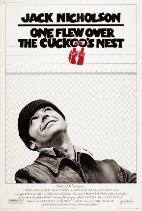
One of my favorite more serious films is One Flew Over the Cuckoo’s Nest. What does Jack Nicholson’s portrayal of a bad guy hoping for easy served time in a mental institution have to do with medical imaging? Well it all starts with the lobotomy. Not to spoil the story, suffice it to say that the movie broaches the topic of lobotomies and how ridiculous they were. Lobotomy was a form of neurosurgery that involved damaging the prefrontal cortex in order to “calm” certain mentally ill patients. Needless to say the procedure was controversial from the beginning (1935 to the early 1970’s) but the author of the discovery, Egas Moniz, was awarded the Nobel Prize in 1949. Maybe not the most sound of decisions by the committee. However, for the time, it was considered progress in a very challenging area of medicine – mental illness. 


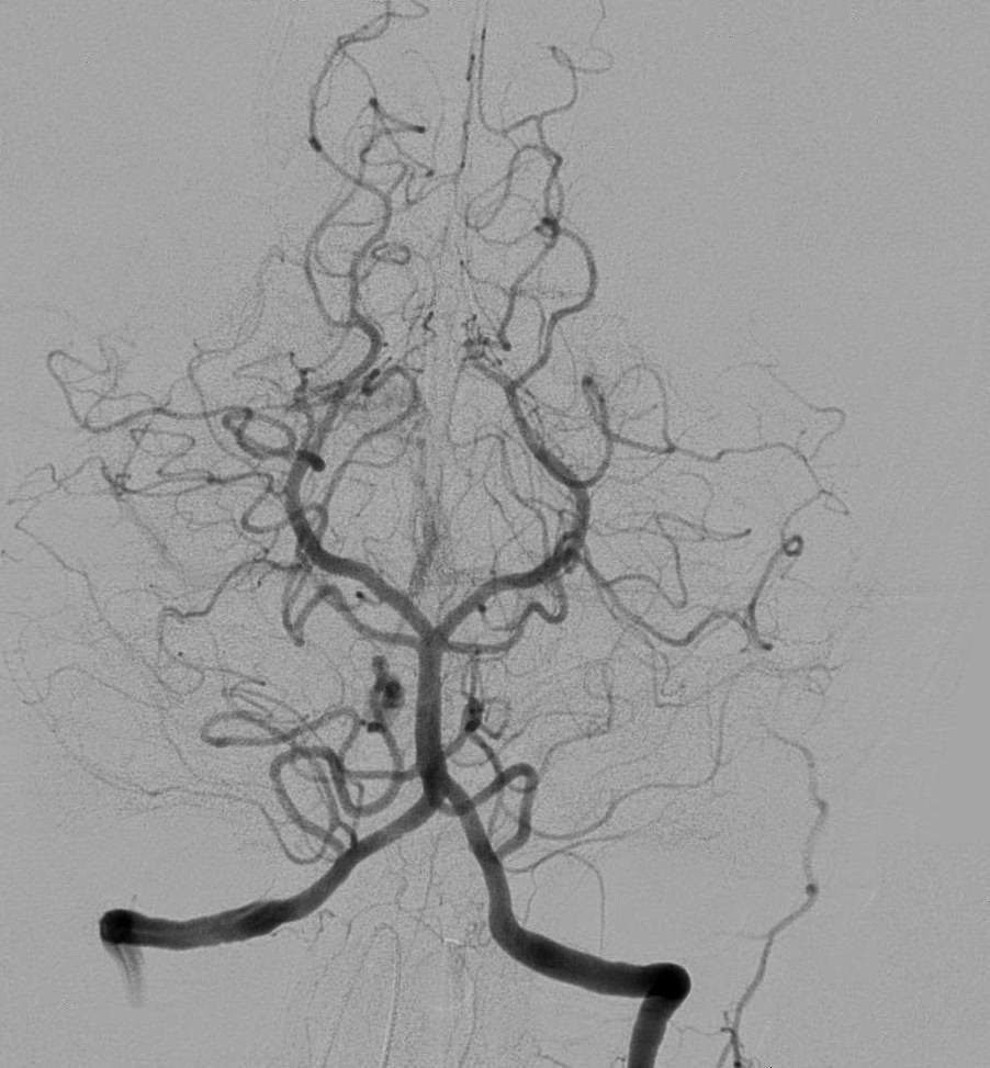
OK, medical imaging? Well as it turns out Moniz (do not confuse with St-Moriz, ahhh skiing…) is also known for developing cerebral angiography – a technique allowing the visualization of blood vessels in and around the brain. 


Moniz was interested in finding a non-toxic substance that would be eliminated from the body, but would not be diluted by the flow of blood before the x-ray could be taken. Another requirement is that the substance could not cause an emboli or clot as this would be a bad thing. Moniz played with salts of iodine and bromine and settled on iodine because of its greater radiographic density. And voila, birth of iodinated radiocontrast agents still in use today. Cool.


Supposedly it took him 9 patients to perfect his angiogram technique. Don’t ask about the first 8…

Moral of the story is: lobotomy bad and cerebral angiography good.


Now for the fun part (see the rules here), using Cuckoo in a sentence by the end of the day:

 
Serious: Hey Bob, when I was visiting my aunt in Australia I spied a little bronze cuckoo in her backyard! This could be my “big year“…


Less serious: Someone won a Nobel Prize for developing the lobotomy? Are you cuckoo?
 
 
Listen to Los Lobos (not short for lobotomy but “the wolves” in Spanish) singing La Bamba to decompress and…
 
… I’ll see you in the blogosphere,
 
 
Pascal Tyrrell

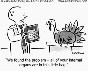
Another successful diagnosis thanks to the x-ray…

For more info see Free Birds

Happy Canadian Thanksgiving!!!

A traditional holiday – originating from the native peoples of the Americas – to celebrate the completion and bounty of the harvest. Well, no harvest for me but I will take the time to appreciate some of the successes of our MiVIP program and this blog over the long weekend.


Thanks for being a part of it!


See you in the blogosphere,

Pascal Tyrrell

Starting a new research project? Think Zorro!

Well, OK maybe think Zotero. The Mask of Zorro was such a great movie I could not resist. Having said that, when starting a new research project it may be helpful for you to think of yourself as Zorro. It may give you that extra zip required to get you through the inevitable research project doldrums…


So what is this Zotero thing anyway? Well Zotero is an open source reference management software that can act as your personal research assistant – helping you to organize and cite the numerous articles that you will be reviewing.


I was talking to Ori the other day – who is in the Radiation Therapy program at the Michener Institute – and he is in the process of planning a research project. As it turns out he has been a member of the MiVIP family since the beginning so he is well aware of my earlier posts that will help him along:


1- Thoughts on how to become a researcher


2- What is in a research question?


3- What makes up a good research question with the F.I.N.E.R. series.


Now how about the reference management software thing? Well, I give you an easy, fun, and instructional e-learning module to help you along. Our group has just finished our first kick at the can (so to speak) and so I invite you to have a look. Here is the link:


MiEducation Zotero e-learning module


Tell us what you think by posting comments and suggestions to this post.


Maybe listen to Ylvis in What Does the Zorro Say? while you go through the module. Fox in spanish is zorro…

 
… and I’ll see you in the blogosphere.
 
 
Pascal Tyrrell


MiWord of the Day Is… Fluoroscope!

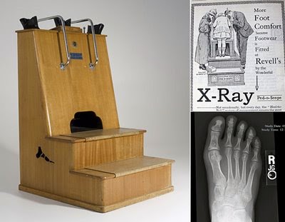
It is hard to believe that the fluoroscope (essentially an x-ray machine used to produce real-time moving images viewed on a screen of the internal structures of a patient) was used to “help” better fit shoes to your feet! From the 1920 to about 1970 you were able to irradiate your feet with x-rays in order to see if you had enough “wiggle-room” in your new shoes! Crazy. 
 
So, the whole concept of Fluroscopy dates back to you know who, Wilhelm Röntgen. We chatted about him here in our blog. He is also responsible for discovering the interesting phenomenon of barium salts fluorescing when exposed to x-rays (see here in our blog). 
 
Basic function of a fluoroscope
Soon after Rontgen’s discovery was announced, Thomas Edison (the light bulb guy) decided he could improve on this whole x-ray thing as these rays were produced by a “glass tube apparatus” – something he knew a lot about. After setting his team to work – he had a team as he was a very successful man in those days following his 1879 patent of the light bulb – they soon discovered the risks of working with x-rays. Edison decided to remove himself (literally!) from x-ray research. But before he did he developed one of the first (and arguably the most advanced in it’s time ) fluoroscopes along with a full line of x-ray kits. He also coined the term “Fluoroscope”. Interesting man…
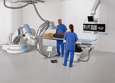
Fluoroscopes have come a long way over the years and are still used today in areas such as orthopedic surgery, gastrointestinal investigations, and angiography but, of course, the dose of x-rays a patient receives is minimized and closely monitored. Have a look at this machine from Siemen’s. “Beam me up Scotty!”. 
So how did all of these machines suddenly flood the shoe retail industry? Good question. As it happens, following the development of the high vacuum, hot cathode, tungsten-target x-ray tube by William Coolidge in 1913 the interest for a portable and reliable machine increased dramatically with the advent of the First World War. The successful deployment of numerous machines during the war to aid army physicians spurred the manufacturing industry to mass produce them. After the war, the impact the fluoroscope had on army medicine flowed into community practice. 
 
Due to the enormous supply of portable x-ray machines at the time following the end of the war, Dr Jacob Lowe introduced the idea of using a modified portable x-ray machine in the shoe retail industry. Voila, fried feet fricassee for the next 50 years!

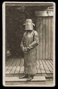
Now If were to be interested in using a fluoroscope to look at my feet I may be inclined to use a suit like this gentleman below is sporting…

WW I x-ray protection suit

Now for the fun part, using Fluoroscope in a sentence by the end of the day:


Serious: Bob, did you know that the foot-o-scope was a modified fluoroscope used to view ones feet when fitting new shoes which delivered on average 13 Roentgens for every 20 second exposure?


Less serious: I heard grampa grumbling he can never find shoes that fit right anymore since they banned fluoroscopes in shoe stores. What is a fluoroscope mommy?


Listen to High Heels to decompress and I’ll see you in the blogosphere.


Pascal Tyrrell

MiWord of the Day Is… Radio!

Easy one today! I thought I would give everyone a break as you have all been working very hard on the MiWord of the day in the past weeks. 


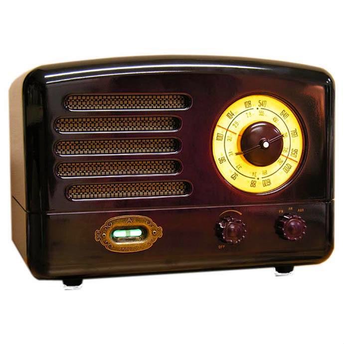
So, what does radio have to do with medical imaging? What a great question! The origin of the root word “Radio” is radiant energy. The radio you immediately think of is the one that is attached to your ear most of the time and has a DJ who selects music to play for your entertainment – along with ads to pay for the station’s bills! The use of “radio” to describe this form of wireless communication comes from the word radiotelegraphy. 


How about if we were simply interested in a medical picture produced by radiant energy? Well you would end up with a radiograph AKA an x-ray! We talked about that word here. Do you see the trend? How about a picture produced by radiant energy in the visible light range of the electromagnetic spectrum? A photograph. Cool.


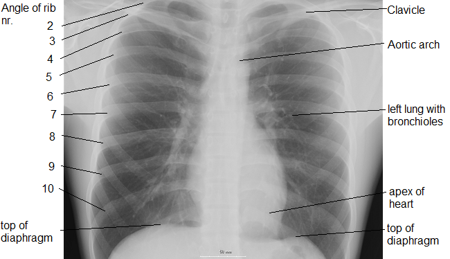
OK now suppose you are an MD working in the emergency department and someone presents with a lung disorder. What do you do? Generally, you order a chest radiograph. As you zap your patient with x-rays you expect that most of them will pass through the chest area – that is mostly filled with air – unchecked and will proceed to expose the film (or trigger the detector) resulting in a dark area. However, if the lungs become filled with abnormal substances more of the x-rays are blocked and result in a lighter (whiter) radiograph. What would you be looking for?


1- Pus – a combination of bacteria and white blood cells as seen with pneumonia.
2- Edema – fluid that leaks into the lungs as seen with heart failure.
3- Hemorrhage – bleeding into the lung cavity as seen with trauma.
4- a solid mass – as seen in lung cancer.


Today, we have to use “Radio” in a sentence (see rules here). Easy! Here are two examples to help you along:


Serious: Bob, you will need to remove your radio from your person before entering the MRI. No metal objects are permissible in the room.


Less serious: I went for a radiograph today and all they did was have me stand in a room by myself and that was it! What a relief. I thought for a moment I was scheduled for a radio-graft…!


Have a listen to my favorite Radiohead to decompress and…


… I’ll see you in the blogosphere,


Pascal Tyrrell