Well, I think it was inevitable. My data science lab has slowly crossed over to the dark side into the world of Machine Learning and Artificial Intelligence.
Let me apologize for being MIA for so long. Life has been pretty hectic these past months as I have been building the MiDATA program here in the Department of Medical Imaging at the University of Toronto. The good news is that the MiVIP program will now be inviting students to participate in machine learning and artificial intelligence in medical image research.
This summer will include the launch our our MiStats+ML program where we will have students from the department of statistical sciences, computer sciences, and life sciences all work together on ML/AI projects in the MiDATA lab.
Stay tuned as we ramp up and get back to some our previous threads like MiWORD of the day…
See you in the blogosphere,
Pascal
“Hour of Code… Part Deux” at the University of Toronto
What hour of code? What code?
The Hour of Code is a global movement reaching tens of millions of students in 180+ countries. The purpose is to to demystify “code”, to show that anybody can learn the basics, and to broaden participation in the field of computer science. Please see here for more info.
I belong to Code.org a non-profit dedicated to expanding access to computer science, and increasing participation by women and underrepresented students of color.
What is the purpose of this event?
To engage young minds and help them see the exciting possibilities computer programming can offer them in their future careers.
Who is coming?
Over 100 students (grades 7 to 11) from Central Toronto Academy (TDSB), St Francis Assisi and St Ignatius of Loyola (TCDSB).
Who will be engaging them (so far)?
University of Toronto:
MiDATA (Data Science unit from the Department of Medical Imaging)
*Prof Pascal Tyrrell – Director, Data Science
*Prof Anne Martel – Medical Biophysics (Machine Learning)
*Dr Mariam Afshin – Research Physicist
Department of Medical Imaging
*Dr Alan Moody – Radiologist and Chair of the Department
Department of Statistical Sciences
*Prof Paul Corey – Senior Biostatistician
*Chris Meaney – Biostatistician
Department of Computer Science
*Prof Steve Engels
Industry:
IBM Watson Health and Merge Healthcare
*Aditya Sriram – Developer, IBM Watson Health Imaging
Microsoft (Big Data and Analytics)
* Sage Franch – Microsoft Canada
SAS Canada
*Mark Morreale – Lead, Academic Program
Community:
Ladies Learning Code – Yaa Otchere
Industry Support:
Google
Tyrrell lab students and Computer Science undergraduates will be acting as ambassadors.
Where and when will the event be held…exactly?
University College, University of Toronto
15 King’s College Circle
Toronto, Ontario
Interested in participating? Contact me at pascal.tyrrell@utoronto.ca!
See you all there,
Pascal Tyrrell
MiDATA hosts “Hour of Code” at the University of Toronto
What hour of code? What code?
The Hour of Code is a global movement reaching tens of millions of students in 180+ countries. The purpose is to to demystify “code”, to show that anybody can learn the basics, and to broaden participation in the field of computer science. Please see here for more info.
I belong to Code.org a non-profit dedicated to expanding access to computer science, and increasing participation by women and underrepresented students of color.
On Wednesday, December 7th at 10AM we will be hosting the inaugural
“MiDATA Hour of Code at UofT”
What is the purpose of this event?
To engage young minds and help them see the exciting possibilities computer programming can offer them in their future careers.
Who is coming?
Over 100 students (grades 7 to 11) from Central Toronto Academy (TDSB), St Francis Assisi and St Ignatius of Loyola (TCDSB).
Who will be engaging them (so far)?
University of Toronto:
MiDATA (Data Science unit from the Department of Medical Imaging)
*Prof Pascal Tyrrell – Director, Data Science
*Prof Anne Martel – Medical Biophysics (Machine Learning)
*John Harvey – Information Architect
MiNE (Medical image Network Enterprise, Sunnybrook Health Science Centre)
*Dr Mariam Afshin – Research Physicist
*Rasha Mahmood – VBIRG
Department of Medical Imaging
*Dr Alan Moody – Radiologist and Chair of the Department
Department of Statistical Sciences
*Prof Jamie Stafford – Statistician and Chair of the Department
*Prof Paul Corey – Senior Biostatistician
Translational Research Program /Institute of Medical Science
*Prof Joseph Ferenbok – Program Director
Department of Computer Science
*Prof Francois Pitt
Faculty of Engineering
*Prof Naomi Matsuura – Department of Materials Science & Engineering
Industry:
IBM Watson Health and Merge Healthcare
*Steve Schudlo – Executive Director, Strategic Alliances, IBM Watson Health Imaging
*Marwan Sati – VP of Development, Clinical Speciality Solutions, Merge Healthcare
*Aditya Sriram – Developer, Watson Health Imaging, IBM
Microsoft (Big Data and Analytics)
*Mark Godfrey – Cloud Architect, TSP – Cloud & Data Center, Microsoft Canada
AceAge
*Spencer Waugh – CEO AceAge
*Sam Campbell – CTO AceAge
*Dylan Horvath – Cortex Design President
SAS Canada
*Mark Morreale – Lead, Academic Program
Community:
Ladies Learning Code – Yaa Otchere
Tyrrell lab students and Computer Science undergraduates will be acting as ambassadors.
Where and when will the event be held…exactly?
University College Media Room (RM 140 and RM148) from 10 AM to 1 PM
University College, University of Toronto
15 King’s College Circle
Toronto, Ontario
Interested in participating? Contact me at pascal.tyrrell@utoronto.ca!
See you all there,
Pascal Tyrrell
The story behind Connectory…
So, you are reading our blog thinking Pascal is a nut – that much is clear – but what of all the students plugged into his group? Are they nuts too?
 Well maybe, but today I am going to talk to you about the group of four (not the group of seven) who started small and grew to be Connectory. John, Maria, Natasha, and Roger met in a graduate course at the University of Toronto and decided to work together on a project about innovation. That’s when they met me, joined “the program”, and got busy! Starting any endeavour from scratch is no easy task. All four had never met before, all came from very different academic backgrounds, and though their initial project was for “credit” the rest was on their own time.
There were some rough times at first but with perseverance comes success and Connectory was born and is just finishing up its first project as a new start-up business. Wow!
Essentially Connectory is a data management solutions software development consulting group that operates in the healthcare space. Check out their webpage here.
Ok, so what? Well this post is not only to congratulate these four on a job well done but also to encourage you to do the same. One thing is for sure: if you don’t try you will not succeed – ever. My programs are all about learning, trying new stuff, benefiting from your successes as well as your failures, and wait for it… giving back. Yup as Uncle Ben said in Spiderman: “With great power comes great responsibility“.
Just wanted to share a good story from our group with you today.
Listen to Bulletproof by La Roux to get pumped and…
… I’ll see you in the blogosphere!
Pascal Tyrrell
The Importance of Research
There’s more to the field of medical imaging than a bunch of stuffy radiologists huddled around a couple of monitors. As I mentioned before in my previous post about the history of the imaging technique, the field has undergone a rapid technological advancement in the past century or so, improving the clinical model of visualization. But let’s take a step back from all the scientific stuff for a brief second and look at these developments in a
slightly different light.
 |
| The SparkNotes illustrated version of this post |
could go a long way. Who knows, you may find yourself presenting your findings at a research symposium, complete with nifty results and statistics to showcase your efforts.
The Key to Research: (Key)Words
Yes, you are not the only one, many people use Google to further explore some of the things they have come across throughout daily encounters. For each instance google is used, whether it be for a song or for neuroscience research and analysis, one thing remains in common: keywords.
Keywords are essential when searching for various types of information, and the options appearing on any search engine are dependent on the keywords given. How does one establish appropriate keywords for a search engine entry?
Faith Balshin
A Crash Course in Medical Imaging
Oddly enough, there’s been a surprising lack of content about medical imaging on a blog with medical imaging in its title. So in order to fill that void, I’ll be providing a brief history on the development of the clinical technique used to visualize the human body.
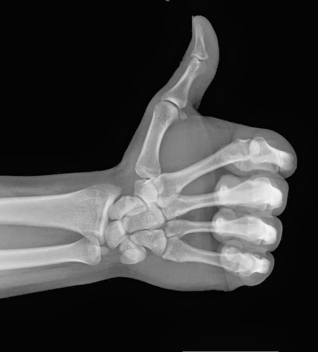
The advent of medical imaging dates all the way back to 1895, following the discovery of X-rays by the German physicist, Wilhelm Conrad Roentgen. The first X-ray picture was then produced, detailing the skeletal composition of his wife’s left hand. However, the actual quality of this imaging process was still very primitive, only allowing for the visualization of bones or foreign objects.
 |
| Much to Dr. Roentgen’s pleasure, Mrs. Roentgen had not discarded her wedding ring |
- Ultrasound – Uses sound waves that are able to penetrate cellular tissue. Once they reflect off the body’s internal organs, the vibrations generate an electrical pulse which can then be reconstructed into an image.
- PET-CT Scan – Positron emission tomography (PET) uses compounds that emit positrons when they decay rather than gamma rays. It is now combined with a computed tomography (CT) device to generate a high-resolution image displaying sectioned layers of the scanned area.
- MRI – A Magnetic Resonance Imaging scanner runs a strong magnetic field through the body, aligning hydrogen protons. As the protons return to their original position in the atom, they generate radio waves, which are then picked up by the scanner and used to create an image based on signal strength.

Fast-forward to present day and over 70 million CT scans, 30 million MRI scans and 2 billion X-rays have been performed worldwide! The field of medical imaging is still growing by the day, with ongoing research leading to new developments.
Thanks for reading,


