Indranil Balki – Summer 2015 ROP at UofT: Another great student experience!

Indranil Balki – ROP Summer 2015


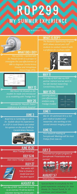
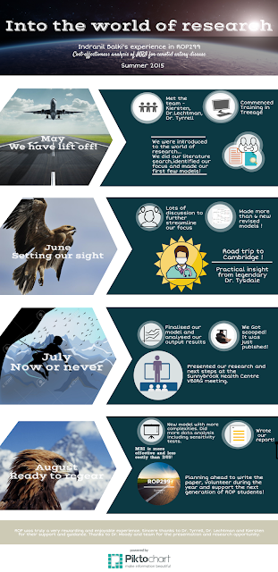
I am one of Dr.Pascal’s 2nd year ROP students at the University of Toronto.  This summer, I had an amazing experience into the realm of research working with my partner Kiersten Thomas, mentor Dr. Eli Lechtman, and supervisor Prof. Pascal Tyrrell. This timeline-cum-infographic highlights some of the especially memorable moments of this journey.
Our main project was focused on constructing a computer simulation to model the effect of choosing either MRI or US as a first line imaging modality to diagnose and treat patients with Carotid Artery Disease. This project involved comprehensive literature search, discussions with experts (including the “Trip to Cambridge”), learning computer software and presentations!
Our team’s models’ main finding was that MRI can be both more effective and no more costly than US as a first line diagnosing tool in measuring carotid artery disease.
 

Thanks to Dr. Pascal for allowing me to share my experience on his blog and I hope you enjoy the read!
 
 
Indranil Balki

An “Egg-cellent” Journey to Investigate Carotid Artery Stenosis in Cambridge, Ontario…


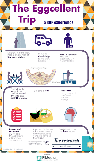
Last Wednesday, my ROP students Kiersten and Indranil, and UofT medical student Eli Lechtman had the opportunity to interview the legendary (see here) Dr. Kim Tysdale – a general practitioner in Cambridge, Ontario.


Why do you ask? Well each year 50,000 Canadians suffer from a stroke with 26% due to carotid artery disease. Carotid artery stenosis is the narrowing of the carotid arteries due to plaque buildup (see atherosclerosis). These plaques can then rupture and create blood clots that travel up to the brain. In turn, these blood clots then get stuck in the brain’s smaller blood vessels, causing a stroke. So plaque = bad and ruptured plaque = worse!


What if we had a clue as to which plaque may rupture? Well, in turns out that the presence of intraplaque hemorrhage (IPH) can help us predict just that! IPH is bleeding within the plaques, which causes them to become more vulnerable (see vulnerable plaque). There is a 6 times greater risk of stroke in people with IPH! And…. a new medical imaging technique called MRIPH imaging allows for visualization of IPH. MRIPH is similar to traditional MRI but highlights the artery walls and looks at the arterial plaques. 


While in Cambridge, Kiersten, Indranil, and Eli presented Dr. Tysdale with an overview of medical imaging techniques for the assessment of carotid artery stenosis with an emphasis on the new MRIPH technique. Informing physicians like Dr. Tysdale which of his patients are more at risk of having vulnerable plaques (by providing information on IPH) could result in patients receiving more appropriate and timely treatment – thereby reducing the number of strokes! 


BTW, we had Dr Tysdale at “hello” (not sure what I am referring to? See here).


So what up with the eggs? Kiersten, Indranil, and Eli also had the chance to tour Dr. Tysdale’s beautiful country house and meet his lovely wife (and nurse!), Eva, who sent everyone home with fresh chicken eggs! Awesome.


Thank you Dr Tysdale for being such a great sport!


Here is their infographic of the trip (great job gang!). Enjoy!

Kiersten Thomas and Indranil Balki – ROP Summer 2015


Stay tuned for more ROP adventures!


Pascal Tyrrell

Research Opportunity Program: Imaging Carotid Stenosis

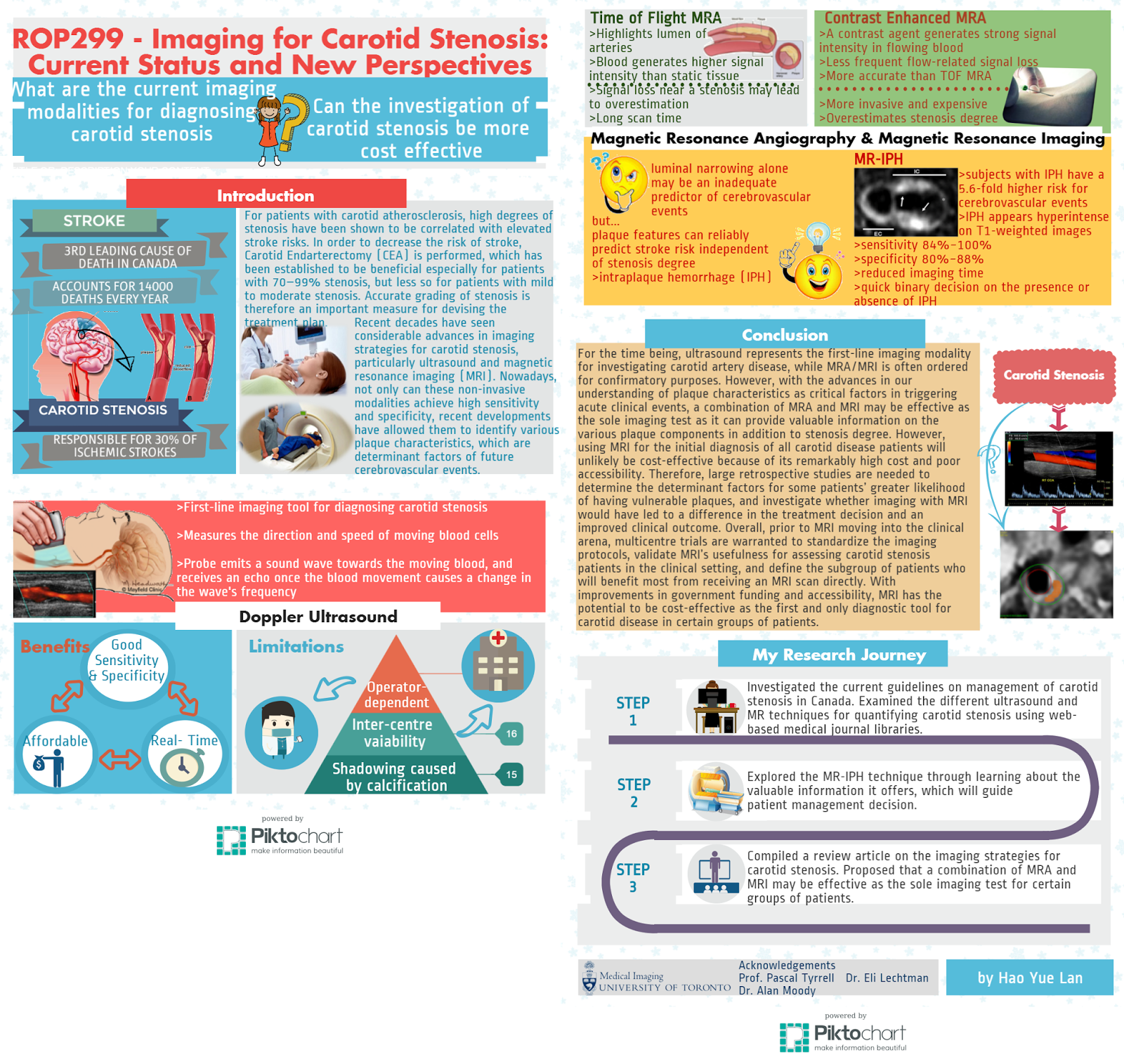
ROP Research Forum March 5th, 2015 – Helena Lan

Helena is a second-year student pursuing a specialist in Pharmacology and Toxicology at the University of Toronto. Participating in ROP299 this past summer (see here) has opened many doors for her. She is currently assisting Pascal on a systematic review on research methodology and biostatistics in medical imaging, working as a research assistant for a medical education study, participating in the research abroad program at Karolinska Institutet this summer… and recently represented us at the March 5th, 2015 ROP research forum!

Helena examined the technical aspects and information provided by two major imaging modalities, MRI and ultrasound, for diagnosing carotid stenosis. She suggests that MRI holds great promise to serve as a cost effective test for carotid stenosis as well as a tool for assessing vessel health and plaque composition that would provide important information for patient management decisions.


Stay tuned, next Sylvia Urbanik will be talking about cost effectiveness…

See you in the blogosphere,

Pascal Tyrrell

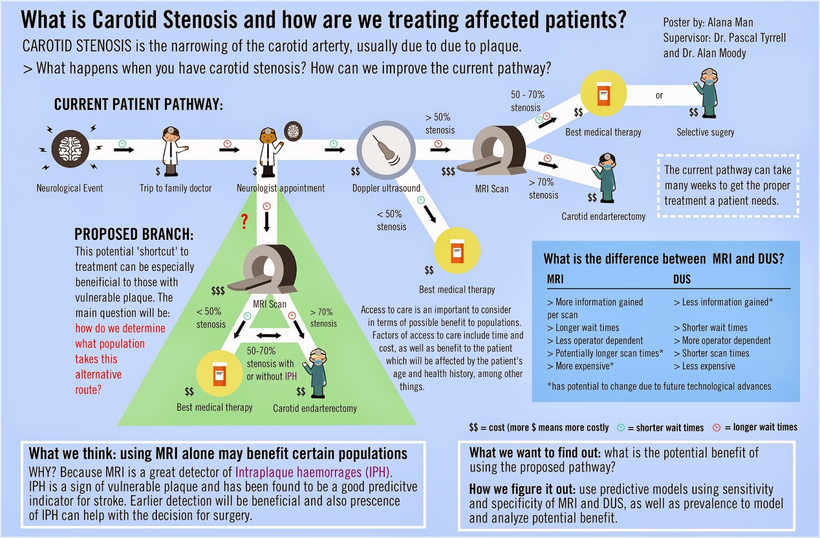
Research Opportunity Program: What Is Carotid Stenosis and How Are We Treating Affected Patients?

ROP Research Forum March 5th, 2015 – Alana Man

Alana Man is a second year University of Toronto student pursuing a specialist in Bioinformatics and Computational Biology and a major in Immunology. She was a Research Opportunity student last summer with MiVIP (see here) and recently represented us at the March 5th, 2015 ROP research forum.

Alana’s project focused on care for patients with carotid stenosis in Toronto, Ontario, Canada. She looked at the different factors contributing to access to diagnostic testing such as procedure costs, wait times, and the differences between available imaging modalities and explores whether MRI could be the diagnostic test of choice over DUS for people at increased risk of carotid stenosis.

Well done, Alana!


Stay tuned, next Helena Lan will be comparing MRI and DUS…


See you in the blogosphere,


Pascal Tyrrell

Helena Lan: Summer 2014 ROP

Helena Lan Summer 2014 ROP

What is research like? If you had asked me this
question several months ago, I would have answered, “You wear a lab coat and
goggles while mixing chemicals or observing organisms. Hopefully something
interesting will happen, so that you get to publish your findings!” Well, after
participating in the Research Opportunity Program (ROP) at the University of
Toronto, I discovered that medical imaging research is more than just
pipetting, and is all the more exciting!

 
So what kind of research is conducted in the medical imaging world? For my ROP, the objective of my project was to evaluate the roles of the non-invasive imaging modalities for diagnosing carotid stenosis. Hence, I engaged in online literature research of the various imaging techniques for assessing this disease. In this process, I also learned to use Zotero to manage all my references, which provides an easy way to generate a bibliography (when the software doesn’t crash every time you open it). After gathering all the pertinent information, I then put together a review article suggesting how a change in the current imaging approach could potentially improve clinical outcome. Who knew a report could be compiled without doing the lab grunt work?     
 
Wait, so this is all a radiologist does? Sitting in front of a computer and typing all day? Of course not! During our time at Sunnybrook Hospital, we got the chance to chat with a radiologist and discovered that she could decide whether patients should be released after taking a look at their diagnostic images. Pretty powerful, eh? That’s not all. We also found out how radiologists identified any abnormalities in patients, as we had the opportunity to work with the VesselMass software which allowed for the delineation of the lumen and vessel wall of arteries on MRI images. Oh, and did I mention we observed an MRI and an ultrasound examination of the carotid arteries, and even got to perform an ultrasound scan ourselves. Super cool!
 
Still craving for more of my ROP experience? Check out my timeline infographic! You will find all the things I learned and all the fun I had there. Last but not least, I’d like to shout out a big THANK YOU to Prof. Pascal Tyrrell and Dr. Eli Lechtman, who guided us every step of the
way. Also, I’m very grateful to Dr. Alan Moody for including us in his research program at Sunnybrook, as well as other members of the VBIRG group who gave us the chance to
participate in various activities. My summer would not have been this fun and meaningful without all of your help!
 
Have fun researching,


Helena Lan