MiWord of the Day Is… PET scan!


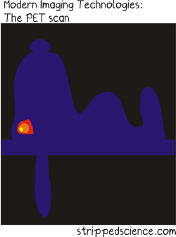
Peanuts. What a great story. The most popular and influential comic strip in history. Snoopy was my first stuffed animal growing up. He still lives with my parents. So what is a PET scan anyway? I don’t recall ever seeing the picture above in any of the Peanuts cartoon strips.


Positron emission tomography (PET) is somewhat of a special medical imaging modality in that it brings together two different technologies from different times. Let me explain. Back in the early 1930s, George Hevesy was a young Hungarian physicist who developed biologically safe and useful radioactive tracers that could be ingested or incorporated into the body in some way.  Physicians would then manually locate where these radioactive tracers had gone in the body by using a Geiger counter at first and then later using special cameras (Kuhl‘s photoscan) to produce a crude emission image. 


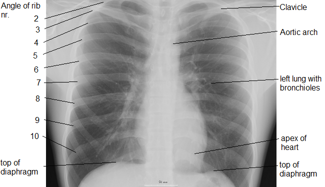
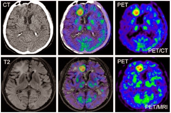
So, how do we get cool pictures like these ones?  Well we would have to wait another 25 years after the development of radioactive tracers by Hevesy for the start of construction of instruments able to not only detect these radioactive sources in the body but to produce tomographic pictures.


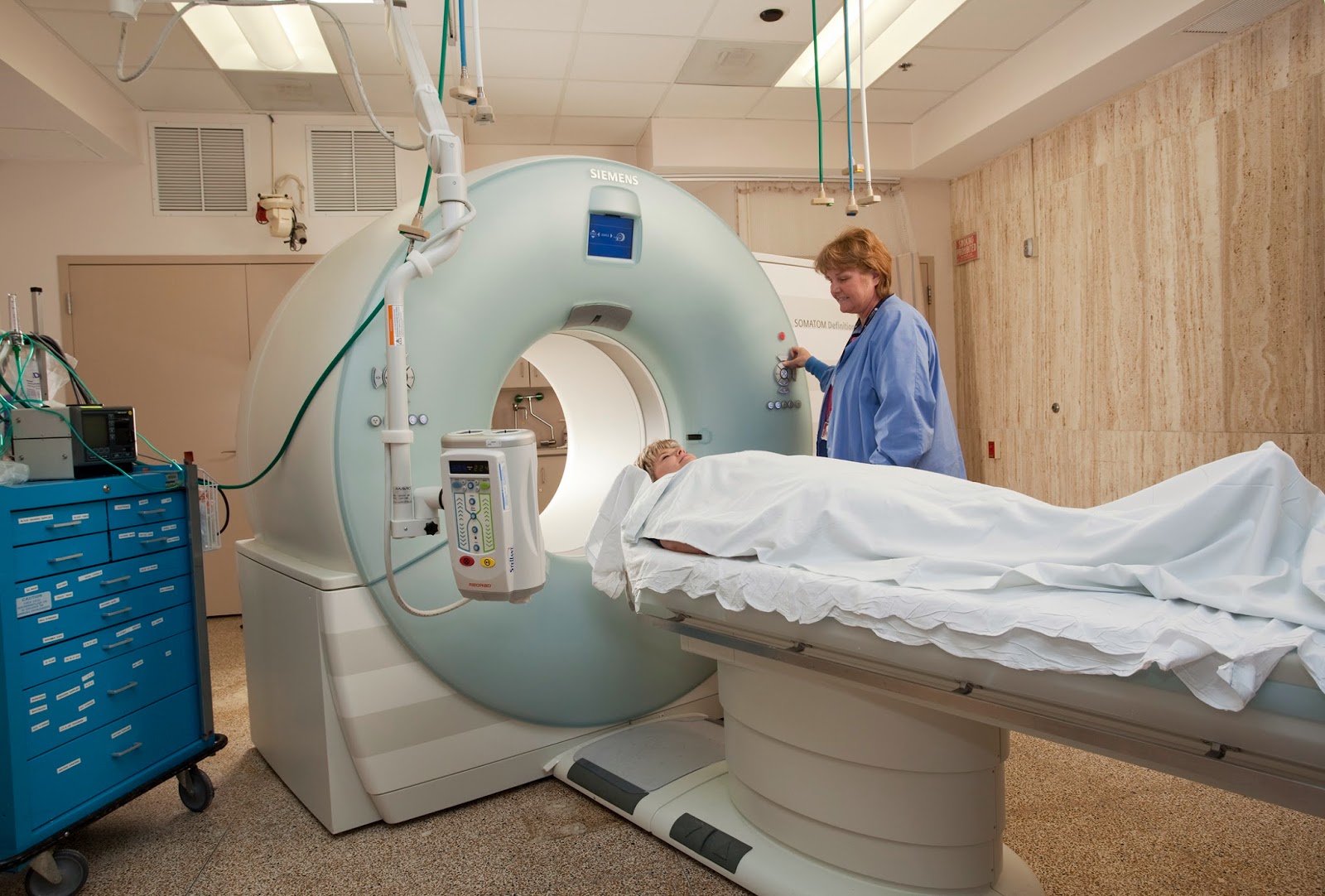
It won’t be until the mid 1970s that PET – as we know it today – would be born. Essentially, a patient receives a emissions scan (PET) and a CT (we talked about that here) or MRI (we talked about that here) scan at the same time. The two scans are then merged together thanks to highly specialized computers (see the pictures in the middle frames). Voila! PET. 


PET is both a medical and research tool. Most often used in clinical oncology (medical imaging of tumors and the search for metastases), it is also important in clinical diagnosis of certain diffuse brain diseases such Alzheimer’s disease and other types of dementia.


Relax your brain a little listening to Radioactive by Imagine Dragons and don’t forget the fun part (see the rules here), using PET scan in a sentence by the end of the day:

Serious: Hey Bob, did you know that much of the success of the PET scan is due to the development of the radiopharmaceutical FDG (deoxyglucose) that lead the way to the characterization of Parkinson’s and Huntington’s disease?

Less serious: I can’t believe they developed yet another PET scan. Wasn’t the CAT scan enough?

See you in the blogosphere,

Pascal Tyrrell


Walk Like an Egyptian!

So, in my last post I talked a little about Mesopotamian medicine (see here). I am certain many of you were thinking: “What? Should he not be talking about ancient Egypt?”. Well, of course, you are right – kind of… 


Egypt rose under the pharaohs during the same period as the Mesopotamian kingdoms (from about 3000 BC). They were known for their crazy ambition and technological prowess. Their medicine was very similar to that of the Mesopotamians in that it was influenced strongly by superstition  and religious beliefs. They too had three types of healers: the swnu who practiced medicine, and, of course, the priests and the sorcerers…


One of the reasons that ancient Egyptian medicine had a greater influence on modern medicine was that they were very good at documenting and archiving their work. The Ebers papyrus (c. 1550 BC) was their principal medical document that measured over 20 meters long (it is a scroll after all) and is the oldest surviving medical book. 


The Egyptians believed we were all born healthy but were susceptible to disorders caused by demons or by intestinal putrefaction. So the importance of eating your fruits and veggies was started way long ago! They also compared our vascular network to that of the River Nile and its canals and, therefore, it was important to keep the flow free from obstructions (see here for another interesting comparison!). Though they did not appreciate vascular plaques (atheroma) at the time they had already started to  figure out the importance of a healthy vasculature. Cool! 


As with Mesopotamia, Egypt’s powerful governance created a good environment for organized medical practice. However, because both regimes were highly codified (implying many strict rules based on religion and superstition that did not allow for discussion and experimentation) it will not be until ancient Greece that the roots of modern medicine will take hold.


Dance around your living room (in private if you must) to Walk Like an Egyptian by The Bangles in order to decompress and…


… I’ll see you in the blogosphere.


Pascal Tyrrell

The story behind Connectory…

So, you are reading our blog thinking Pascal is a nut – that much is clear – but what of all the students plugged into his group? Are they nuts too?


Well maybe, but today I am going to talk to you about the group of four (not the group of seven) who started small and grew to be Connectory. John, Maria, Natasha, and Roger met in a graduate course at the University of Toronto and decided to work together on a project about innovation. That’s when they met me, joined “the program”, and got busy! Starting any endeavour from scratch is no easy task. All four had never met before, all came from very different academic backgrounds, and though their initial project was for “credit” the rest was on their own time.


There were some rough times at first but with perseverance comes success and Connectory was born and is just finishing up its first project as a new start-up business. Wow! 


Essentially Connectory is a data management solutions software development consulting group that operates in the healthcare space. Check out their webpage here.


Ok, so what? Well this post is not only to congratulate these four on a job well done but also to encourage you to do the same. One thing is for sure: if you don’t try you will not succeed – ever. My programs are all about learning, trying new stuff, benefiting from your successes as well as your failures, and wait for it… giving back. Yup as Uncle Ben said in Spiderman: “With great power comes great responsibility“. 


Just wanted to share a good story from our group with you today.


Listen to Bulletproof by La Roux to get pumped and…


… I’ll see you in the blogosphere!


Pascal Tyrrell

Who is your neighbor?

Classic Seth Rogan movie. Today we will be talking about good neighbors as a followup to my first post “What cluster Are You From?“. If you want to learn a little about bad neighbors watch the trailer to the movie Neighbors.


So let’s say you are working with a large amount of data that contains many, many variables of interest. In this situation you are most likely working with a multidimensional model. Multivariate analysis will help you make sense of multidimensional space and is simply defined as a situation when your analysis incorporates more than 1 dependent variable (AKA response or outcome variable). 


*** Stats jargon warning*** 
Mulitvariate analysis can include analysis of data covariance structures to better understand or reduce data dimensions (PCA, Factor Analysis, Correspondence Analysis) or the assignment of observations to groups using a unsupervised methodology (Cluster Analysis) or a supervised methodology (K Nearest Neighbor or K-NN). We will be talking about the later today.


*** Stats-reduced safe return here***
Classification is simply the assignment of previously unseen entities (objects such as records) to a class (or category) as accurately as possible.  In our case, you are fortunate to have a training set of entities or objects that have already been labelled or classified and so this methodology is termed “supervised”. Cluster analysis is unsupervised learning and we will talk more about this in a later post.


Let’s say for example you have made a list of all of your friends and labeled each one as “Super Cool”, “Cool”, or “Not cool”. How did you decide? You probably have a bunch of attributes or factors that you considered. If you have many, many attributes this process could be daunting. This is where k nearest neighbor or K-NN comes in. It considers the most similar other items in terms of their attributes, looks at their labels, and gives the unassigned object the majority vote!


This is how it basically works:


1- Defines similarity (or closeness) and then, for a given object, measures how similar are all the labelled objects from your training set. These become the neighbors who each get a vote.


2- Decides on how many neighbors get a vote. This is the k in k-NN.


3- Tallies the votes and voila – a new label! 


All of this is fun but will be made much easier using the k-NN algorithm and your trusty computer!


So, now you have an idea about supervised learning technique that will allow you to work with a multidimensional data set. Cool.


Listen to Frank Sinatra‘s The Girl Next Door to decompress and I’ll see you in the blogosphere…


Pascal Tyrrell

MiWord of the Day Is… CAT scan!

A what scan? I am actually a cat guy myself. Not to say I don’t love dogs but if I had to make a choice…


I just finished reading a fantastic book by David Dosa entitled “Making Rounds With Oscar”. The premise of the book is a story about an extraordinary cat but the subject matter is very serious –  dementia and end-of-life care in the elderly. Have a gander.


So what the heck is a cat scan and what does it have to do with medical imaging? 


CT scans – also referred to as computerized axial tomography (CAT) – are special X-ray tests that produce cross-sectional images of the body using X-rays and a computer. CT was developed independently by a British engineer named Sir Godfrey Hounsfield and Dr. Alan Cormack and were jointly awarded the Nobel Prize in 1979. Yes, more Nobel prize winners…


In a nutshell, x-ray computed tomography:


– uses data from several X-ray images of structures inside the body and converts them into 3D pictures – especially useful for soft tissues.


– emits a series of narrow beams through the human body, producing more detail than standard single beam X-rays.


– is able to distinguish tissues inside a solid organ. A CT scan is able to illustrate organ tear and organ injury quickly and so is often used for accident victims.


– is analyzed by radiologists.


Unfortunately, unlike MRI scans, a CT scan uses X-rays and therefore are a source of ionizing radiation.


Now for the fun part (see the rules here), using CAT Scan in a sentence by the end of the day:

Serious: Hey Bob, did you know that the recorded image of a CAT Scan is called a tomogram? 

Less serious: My GP suggested that howling at the moon at night is not normal behavior and he wants to send me for a CAT scan. What? No way, I’m allergic to cats…

OK, listen to Cat Stevens to decompress and I’ll see you in the blogosphere…


Pascal Tyrrell

MiWord of the Day Is… Cinemaradiology!

Yes, it is Halloween today and my kids could barely contain themselves getting ready for school. I suspect today will not be very productive as they count down the minutes before heading out to terrorize my neighbors.


Anyway, how about this for a scary thought: cinemaradiology! In the late 1800’s John MacIntyre at the Gasgow Royal Infirmary experimented with producing X-ray motion pictures. What!!!? He tried exposing film by passing it between the screen of the fluoroscope and the x-ray tube and by simply filming the fluoroscopic screen. This latter method was very difficult because, as all of you budding radiologists know, the images viewed on the fluoroscope screen were dim and of poor resolution at the time.


For years researchers worked on perfecting cinemaradiology. However, during those early years of discovery they lost interest when they realized that sharper images were possible when BOTH patients and investigators were exposed together AND that excessive radiation was a bad thing – duh! 


It would only be many many years later that fluoroscope screen technology would be improved to allow for brighter and higher resolution images (and without frying the patient and everyone around!). 

Fluoroscopy is a study of moving body structures – similar to an x-ray “movie.”  A continuous x-ray beam is passed through the body part being examined, and is transmitted to a TV-like monitor so that the body part and its motion can be seen in detail. As an imaging tool, Fluoroscopy is used in many types of examinations and procedures.

To my knowledge, no actors from the cinemaradiology era ever became successful stars in Hollywood…


No need to use the MiWord of the day in a sentence today (see rules here) as I realize you are busy getting ready for Halloween and need a break!


Decompress listening to the classic song Thriller by the King of Pop Michael Jackson and I’ll see you in the blogosphere…


Pascal Tyrrell


Another successful diagnosis thanks to the x-ray…

For more info see Free Birds

Happy Canadian Thanksgiving!!!

A traditional holiday – originating from the native peoples of the Americas – to celebrate the completion and bounty of the harvest. Well, no harvest for me but I will take the time to appreciate some of the successes of our MiVIP program and this blog over the long weekend.


Thanks for being a part of it!


See you in the blogosphere,

Pascal Tyrrell

Starting a new research project? Think Zorro!

Well, OK maybe think Zotero. The Mask of Zorro was such a great movie I could not resist. Having said that, when starting a new research project it may be helpful for you to think of yourself as Zorro. It may give you that extra zip required to get you through the inevitable research project doldrums…


So what is this Zotero thing anyway? Well Zotero is an open source reference management software that can act as your personal research assistant – helping you to organize and cite the numerous articles that you will be reviewing.


I was talking to Ori the other day – who is in the Radiation Therapy program at the Michener Institute – and he is in the process of planning a research project. As it turns out he has been a member of the MiVIP family since the beginning so he is well aware of my earlier posts that will help him along:


1- Thoughts on how to become a researcher


2- What is in a research question?


3- What makes up a good research question with the F.I.N.E.R. series.


Now how about the reference management software thing? Well, I give you an easy, fun, and instructional e-learning module to help you along. Our group has just finished our first kick at the can (so to speak) and so I invite you to have a look. Here is the link:


MiEducation Zotero e-learning module


Tell us what you think by posting comments and suggestions to this post.


Maybe listen to Ylvis in What Does the Zorro Say? while you go through the module. Fox in spanish is zorro…

 
… and I’ll see you in the blogosphere.
 
 
Pascal Tyrrell


MiWord of the Day Is… Radio!

Easy one today! I thought I would give everyone a break as you have all been working very hard on the MiWord of the day in the past weeks. 


So, what does radio have to do with medical imaging? What a great question! The origin of the root word “Radio” is radiant energy. The radio you immediately think of is the one that is attached to your ear most of the time and has a DJ who selects music to play for your entertainment – along with ads to pay for the station’s bills! The use of “radio” to describe this form of wireless communication comes from the word radiotelegraphy


How about if we were simply interested in a medical picture produced by radiant energy? Well you would end up with a radiograph AKA an x-ray! We talked about that word here. Do you see the trend? How about a picture produced by radiant energy in the visible light range of the electromagnetic spectrum? A photograph. Cool.


OK now suppose you are an MD working in the emergency department and someone presents with a lung disorder. What do you do? Generally, you order a chest radiograph. As you zap your patient with x-rays you expect that most of them will pass through the chest area – that is mostly filled with air – unchecked and will proceed to expose the film (or trigger the detector) resulting in a dark area. However, if the lungs become filled with abnormal substances more of the x-rays are blocked and result in a lighter (whiter) radiograph. What would you be looking for?


1- Pus – a combination of bacteria and white blood cells as seen with pneumonia.
2- Edema – fluid that leaks into the lungs as seen with heart failure.
3- Hemorrhage – bleeding into the lung cavity as seen with trauma.
4- a solid mass – as seen in lung cancer.


Today, we have to use “Radio” in a sentence (see rules here). Easy! Here are two examples to help you along:


Serious: Bob, you will need to remove your radio from your person before entering the MRI. No metal objects are permissible in the room.


Less serious: I went for a radiograph today and all they did was have me stand in a room by myself and that was it! What a relief. I thought for a moment I was scheduled for a radio-graft…!


Have a listen to my favorite Radiohead to decompress and…


… I’ll see you in the blogosphere,


Pascal Tyrrell

MiWord of the Day Is… Ionizing!

So what the heck is ionizing radiation? Well consider the following conundrum about x-rays. They can:


1- cause cancer
2- be used to detect cancer.
3- be used to treat cancer.


Whaaat? How does that work?  We use the term ionizing  when the radiation has sufficient energy to detach electrons from molecules causing them to become chemically reactive ions


The name atom means “indivisable” and are incredibly small. They are made up of protons, neutrons, and electrons with about 99.9% of its mass concentrated in the nucleus that holds a positive charge. A surrounding negatively charged cloud of electrons makes up the difference and the atom stays together due to the attraction between the two. 


OK, so here is the rub: if an atom gains or loses an electron it becomes an ion and generally results in a very chemically reactive substance. This process to produce an ion can be achieved by many ways but one of the most important is electromagnetic radiation (we’ve talked about this already here). Radioactive materials such as radium emit ionizing radiation as does x-ray tubes. There is even such a thing as cosmic radiation (Yup, we talked about that here!). 


Now x-rays produce photons which are the same particles that make up visible light but at a much shorter wavelength and higher energy. When they penetrate through a solid object they will most often simply pass through. However, if they pass by close enough to an electron they can transfer their energy and in the process knock it out orbit producing an ion. Also, the more dense the object the more often the photons are blocked from travelling through resulting in a differential effect on a film or sensor placed on the opposite side. This is how we are able to see inside the body using x-rays.


The problem about ionizing radiation is that the resulting chemically reactive ions can result in DNA damage. Often the cell can repair itself resulting in no permanent damage. Other times, however, permanent damage occurs and can result in cell death (a good thing if they are cancerous cells) or DNA mutations that can in turn lead to the promotion of cancer – bummer.


Now on to using ionizing in a sentence today (not sure about the rules? See here):
Serious example – Bob, don’t stand too close to the x-ray machine. You wouldn’t want to be exposed to ionizing radiation that could damage the DNA in your cells…


Less serious – You wouldn’t believe what happened to me at work today! I was at the photocopy machine getting ready to change the toner cartridge and Bill from sales said:”Let me do that for you, Honey”. He is so ionizing or patronizing or whatever. He makes me mad… 


Listen to Just Because by Raygun to get ready for the weekend and I’ll see you back in the blogosphere soon.


Pascal Tyrrell