MiWord of the Day Is… CAT scan!

A what scan? I am actually a cat guy myself. Not to say I don’t love dogs but if I had to make a choice…


I just finished reading a fantastic book by David Dosa entitled “Making Rounds With Oscar”. The premise of the book is a story about an extraordinary cat but the subject matter is very serious –  dementia and end-of-life care in the elderly. Have a gander.


So what the heck is a cat scan and what does it have to do with medical imaging? 
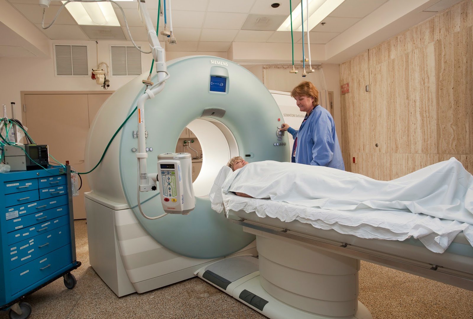

CT scans – also referred to as computerized axial tomography (CAT) – are special X-ray tests that produce cross-sectional images of the body using X-rays and a computer. CT was developed independently by a British engineer named Sir Godfrey Hounsfield and Dr. Alan Cormack and were jointly awarded the Nobel Prize in 1979. Yes, more Nobel prize winners…


In a nutshell, x-ray computed tomography:


– uses data from several X-ray images of structures inside the body and converts them into 3D pictures – especially useful for soft tissues.


– emits a series of narrow beams through the human body, producing more detail than standard single beam X-rays.


– is able to distinguish tissues inside a solid organ. A CT scan is able to illustrate organ tear and organ injury quickly and so is often used for accident victims.


– is analyzed by radiologists.


Unfortunately, unlike MRI scans, a CT scan uses X-rays and therefore are a source of ionizing radiation.


Now for the fun part (see the rules here), using CAT Scan in a sentence by the end of the day:

Serious: Hey Bob, did you know that the recorded image of a CAT Scan is called a tomogram? 

Less serious: My GP suggested that howling at the moon at night is not normal behavior and he wants to send me for a CAT scan. What? No way, I’m allergic to cats…

OK, listen to Cat Stevens to decompress and I’ll see you in the blogosphere…


Pascal Tyrrell

MiWord of the Day Is… Fluoroscope!

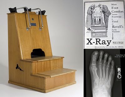
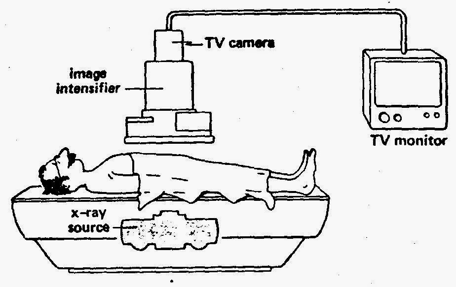
It is hard to believe that the fluoroscope (essentially an x-ray machine used to produce real-time moving images viewed on a screen of the internal structures of a patient) was used to “help” better fit shoes to your feet! From the 1920 to about 1970 you were able to irradiate your feet with x-rays in order to see if you had enough “wiggle-room” in your new shoes! Crazy. 
 
So, the whole concept of Fluroscopy dates back to you know who, Wilhelm Röntgen. We chatted about him here in our blog. He is also responsible for discovering the interesting phenomenon of barium salts fluorescing when exposed to x-rays (see here in our blog). 
 
Basic function of a fluoroscope
Soon after Rontgen’s discovery was announced, Thomas Edison (the light bulb guy) decided he could improve on this whole x-ray thing as these rays were produced by a “glass tube apparatus” – something he knew a lot about. After setting his team to work – he had a team as he was a very successful man in those days following his 1879 patent of the light bulb – they soon discovered the risks of working with x-rays. Edison decided to remove himself (literally!) from x-ray research. But before he did he developed one of the first (and arguably the most advanced in it’s time ) fluoroscopes along with a full line of x-ray kits. He also coined the term “Fluoroscope”. Interesting man…
Fluoroscopes have come a long way over the years and are still used today in areas such as orthopedic surgery, gastrointestinal investigations, and angiography but, of course, the dose of x-rays a patient receives is minimized and closely monitored. Have a look at this machine from Siemen’s. “Beam me up Scotty!”. 
So how did all of these machines suddenly flood the shoe retail industry? Good question. As it happens, following the development of the high vacuum, hot cathode, tungsten-target x-ray tube by William Coolidge in 1913 the interest for a portable and reliable machine increased dramatically with the advent of the First World War. The successful deployment of numerous machines during the war to aid army physicians spurred the manufacturing industry to mass produce them. After the war, the impact the fluoroscope had on army medicine flowed into community practice. 
 
Due to the enormous supply of portable x-ray machines at the time following the end of the war, Dr Jacob Lowe introduced the idea of using a modified portable x-ray machine in the shoe retail industry. Voila, fried feet fricassee for the next 50 years!

Now If were to be interested in using a fluoroscope to look at my feet I may be inclined to use a suit like this gentleman below is sporting…

WW I x-ray protection suit

Now for the fun part, using Fluoroscope in a sentence by the end of the day:


Serious: Bob, did you know that the foot-o-scope was a modified fluoroscope used to view ones feet when fitting new shoes which delivered on average 13 Roentgens for every 20 second exposure?


Less serious: I heard grampa grumbling he can never find shoes that fit right anymore since they banned fluoroscopes in shoe stores. What is a fluoroscope mommy?


Listen to High Heels to decompress and I’ll see you in the blogosphere.


Pascal Tyrrell

MiWord of the Day Is… Ionizing!

So what the heck is ionizing radiation? Well consider the following conundrum about x-rays. They can:


1- cause cancer
2- be used to detect cancer.
3- be used to treat cancer.


Whaaat? How does that work?  We use the term ionizing  when the radiation has sufficient energy to detach electrons from molecules causing them to become chemically reactive ions


The name atom means “indivisable” and are incredibly small. They are made up of protons, neutrons, and electrons with about 99.9% of its mass concentrated in the nucleus that holds a positive charge. A surrounding negatively charged cloud of electrons makes up the difference and the atom stays together due to the attraction between the two. 


OK, so here is the rub: if an atom gains or loses an electron it becomes an ion and generally results in a very chemically reactive substance. This process to produce an ion can be achieved by many ways but one of the most important is electromagnetic radiation (we’ve talked about this already here). Radioactive materials such as radium emit ionizing radiation as does x-ray tubes. There is even such a thing as cosmic radiation (Yup, we talked about that here!). 


Now x-rays produce photons which are the same particles that make up visible light but at a much shorter wavelength and higher energy. When they penetrate through a solid object they will most often simply pass through. However, if they pass by close enough to an electron they can transfer their energy and in the process knock it out orbit producing an ion. Also, the more dense the object the more often the photons are blocked from travelling through resulting in a differential effect on a film or sensor placed on the opposite side. This is how we are able to see inside the body using x-rays.


The problem about ionizing radiation is that the resulting chemically reactive ions can result in DNA damage. Often the cell can repair itself resulting in no permanent damage. Other times, however, permanent damage occurs and can result in cell death (a good thing if they are cancerous cells) or DNA mutations that can in turn lead to the promotion of cancer – bummer.


Now on to using ionizing in a sentence today (not sure about the rules? See here):
Serious example – Bob, don’t stand too close to the x-ray machine. You wouldn’t want to be exposed to ionizing radiation that could damage the DNA in your cells…


Less serious – You wouldn’t believe what happened to me at work today! I was at the photocopy machine getting ready to change the toner cartridge and Bill from sales said:”Let me do that for you, Honey”. He is so ionizing or patronizing or whatever. He makes me mad… 


Listen to Just Because by Raygun to get ready for the weekend and I’ll see you back in the blogosphere soon.


Pascal Tyrrell