Michener Institute Series: Princess Margaret Hospital, Toronto, Ontario


As first year Radiation Thearpy students here a The Michener Institute, we are currently in our 4th week of clinical placements! As promised, here’s a little update about the experiences Jennifer and Ori are going through at Princess Margaret Hospital.

Jennifer: I’ve been placed in Unit 10 which specializes in treating patients with Genitourinary, Gynae and Lower Gastrointestinal cancer.

Ori: I’m on Unit 14 and we treat breast cancer and palliative cancers.

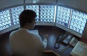
We are proud to say that we are enjoying our experience here. Our duty as students in training is to follow the radiation therapist and learn what they do. The job of a therapist is to treat cancer using a machine called Linear Accelerator (Linac) to deliver ionizing radiation. Patients will typically come once a day for the next couple of weeks, so we see the same patients every day and therefore really get to know our patients well. There is a fair amount of patient interaction, which is one of our favorite parts of the job. Along with patient interactions, we also get to use the equipment, which mainly includes operating the Linac machine (the machine that delivers the radiation) and taking X-rays or CT scans to make sure the patient is in the right position. Every day is a new experience and we are constantly learning new skills. We get a better insight of the patient’s perspective during the entire span of their radiation treatment. For example most patients in unit 10 are required to have a full bladder and empty rectum. Having to hold their pee can be quite difficult for some patients, especially when there are delays, which pushes Unit 10 to be a very fast paced environment. Overall our first 4 weeks of clinical has been an exceptionally valuable experience and we’re looking forward to our next 4 weeks!


Until next time!

Jennifer and Ori


Michener Institute Series: Clinical Placement Site – Kingston Ontario


(Kingston City Hall)
It has been a month since the start of summer clinical placement, and I am currently
completing my placement in Kingston General Hospital (KGH) here at Kingston, Ontario.  Kingston is a nice beautiful town located at the north side of the entrance of outflow of St Lawrence River from Lake Ontario; it was the
first capital of Canada when Canada was still a province of British colony.
 
KGH host one of the most eastern cancer center in Ontario and it has a beautiful view because it is situated by the side of Lake Ontario, its front entrance open to the water. It is a perfect place for lunch and enjoys the sun during summer time.


      (KGH cancer centre front entrance)    
               
          (MacDonald
Park by water, in front of cancer centre)


The past month was phenomenal, words cannot fully describe the knowledge and experience we gain from clinical practice. The transition from purely academic to hands on
practice is eye-opening and a bit hectic; because each patient is unique and no knowledge from books can prepare you how to interact with all patients.  It is interesting to learn from the therapists, the way they educate patients on their first day of treatment, the type of approach to each patient base on the assessment they do during the conversation with them. It’s amazing how much compassion the therapists have for patients and how much they care for them.
 
 
During the first two weeks in CT simulation unit, I made my first mask and had my own mask made for treatment to head and neck regions. The mask is made of pliable plastics. They come in as a sheet of plastic in a frame, and are put into a warm/hot water bath for 2-4 minutes to makes it pliable, after the mask is taken out of the warm water bath there is a 30-60 seconds window before it hardens. The therapist takes out the mask, tower dry it as much as possible and covers it on patient’s head as fast as possible.  The therapists are very efficient at their job, but what is amazing are the patients going through the process; imagine a warm and moist piece of plastic cover you face, harden in an instant and lock your head into position, and afterword you cannot move for 5-10 minutes for CT scan. I never had thought of the discomfort till I experience it myself.


(My 1st  mask, can kinda see my face print)
 
So far the experience here is amazing, and hopefully the coming June will be equally fantastic as well.
 
Till next time.


Gordon

Repeat After Me…

So, in my last post (Agreement Is Difficult) we started to talk about agreement which measures “closeness” between things.  We saw that agreement is broadly defined by accuracy and precision. Today, I would like to talk a little more about the latter.


 The Food and Drug Administration (FDA) defines precision as “the degree of scatter between a series of measurements obtained from multiple sampling of the same homogeneous sample under the prescribed conditions”. This means precision is only comparable under the same conditions and generally comes in two flavors: 


1- Repeatability which measures the purest form of random error – not influenced by any other factors. The closeness of agreement between measures under the exact same conditions, where “same condition” means that nothing has changed other than the times of the measurements.


2- Reproducibility is similar to repeatability but represents the precision of a given method under all possible conditions on identical subjects over a short period of time. So, same test items but in different laboratories with different operators and using different equipment for example.


Now, when considering agreement if one of the readings you are collecting is an accepted reference then you are most probably interested in validity (we will talk about this a future post) which concerns the interpretation of your measurement. On the other hand if all of your readings are drawn from a common population then you are most likely interested in assessing the precision of the readings – including repeatability and reproducibility.


As we have just seen, not all repeats are the same! Think about what it is that you want to report before you set out to study agreement – or you could be destined to do it over again as does Tom Cruise in his latest movie Edge of Tomorrow where is lives, dies, and then repeats until he gets it right… 


See you in the blogosphere,


Pascal Tyrrell

Agreement Is Difficult: So What Are We Gonna Do? I Dunno, What You Wanna Do?

It is never easy to come to an agreement – even amongst friends! The Vultures from Disney’s The Jungle Book (oldie but a goodie) certainly know this. In medical research measuring agreement is also a challenge. In this series of posts I am going to talk about agreement and how it is measured.


Agreement measures the “closeness” between things. It is a broad term that contains both “accuracy” and “precision”. So, let’s say you are shopping for screen protectors for your wonderful new phone. You head to the internet and start going through the gazillion links advertising screen protectors of all sizes and styles. As you just spent your savings on the phone, you do not have much money left over for the screen protector. You decide on a generic brand and order a pack of 10. After an unbearable wait of a week to receive them in the mail you open the pack and find that even though you ordered the screen protector to fit your specific phone they are a little small… except for two that fit perfectly! What? That’s annoying.


So, how close are the screen protectors to being “true” to the expected product? This is agreement. Now, most of them are a little small. This represents poor “accuracy”. This is because there exists a systematic bias. If you took the mean size of these 10 protectors you would find that it deviated from the true expected value – size in this case. Furthermore, you found that two of the 10 protectors actually fit your phone screen rather well. This is great, but this inconsistency between your protectors represents poor “precision”. This time we are interested in the degree of scatter between your measurements – a measure of within sample variation due to random error (see my Dickens post for more info).


Now the concepts of accuracy and precision originated in the physical sciences where direct measurements are possible. Not to be outdone, the social sciences (and then soon to adopt medical sciences!) decided to establish similar concepts – validity and reliability. We will discuss these in a latter post but for now simply remember that the main differences are that a reference is required for validity and that both validity and reliability are most often assessed with scaled indices.


Phew! That was a little confusing. Have a listen to We Just Disagree from Dave Mason to relax.


Next post we will look a little more closely at two special kinds of precision – repeatability and reproducibility.


See you in the blogoshere,


Pascal Tyrrell

What Makes a F.I.N.E.R. Research Question? F is for…

Fugitive? Well that is certainly how you can feel when out with your friends and knowing you should be home studying for your upcoming stats 101 exam. Not sure how that feels? Watch the trailer from the fantastic movie The Fugitive.


Now that you are well versed in dreaming up research questions and driving everyone around you nuts, I thought I would chat a little about how you can assess whether you have a good one “on the line” or a stinker.


A great mnemonic suggested by Hulley et al is F.I.N.E.R. and suggests that research questions need to be feasible, interesting, novel, ethical, and relevant. Today we will talk about the first one.


F is for FEASIBLE. When you are creating a research question you must always ask yourself: “Can I answer this question?”. There are many reasons that may stop you from completing your quest to answer a given question. Maybe not as dramatic as in the Quest for Camelot, but it is important to take note of your limitations:


1- Do you or other members of your team have the know-how or technical expertise to plan, execute, and analyze the study required to answer your question? Maybe you need to brush up on your skills before embarking on your adventure… or phone a friend!


2- Can you afford the time to complete your quest? Do you have the money to pay for it? Gold wins wars not soldiers (Game of Thrones season 1)! Always plan ahead so that your study does not get compromised or cancelled because you don’t have the money or the time to continue (summer student projects often fall pray to this).


3- Do you have access to enough subjects to appropriately power your study? Sample size calculation is a fun and challenging topic and we will address this in later posts. If you want to know how many of your classmates – say 100 – want to go camping over the weekend to celebrate the end of the school year, how many should you ask before you are confident it will be worth your effort to organize the trip? All of them? Some of them? But how many…


4- Are you asking too much at once? Is the scope of your research question too broad? Focus on the most important goals. 


Don’t become a “Jack of all trades and master of none“! Aim for a better answer to the main question that you are interested in.


Next post we will move on to I…

See you in the blogosphere,


Pascal Tyrrell


The Importance of Research

There’s more to the field of medical imaging than a bunch of stuffy radiologists huddled around a couple of monitors. As I mentioned before in my previous post about the history of the imaging technique, the field has undergone a rapid technological advancement in the past century or so, improving the clinical model of visualization. But let’s take a step back from all the scientific stuff for a brief second and look at these developments in a
slightly different light.

During the early stages of medical imaging, X-rays were able to provide people with an initial view of the internal structure of the human body. As limited as that first view may have been, it still played a pivotal role in both challenging and changing people’s perceptions on the human body – to the point where these details would eventually become common knowledge. Without all the major advancements in medical imaging, we could well expect to still be living in the dark.
To really hammer this point home, further advancements in the field would only continue to build on our understanding. What was once the accepted view of the human body has now been given a complete overhaul, thanks to the availability of imaging devices able to produce higher-resolution cross-sectional pictures.

The SparkNotes illustrated version of this post
So what’s the common thread in all of this? Research, of course. While the idea of research leading to new and exciting developments is a pretty basic concept in and of itself, it’s still an important one to keep in mind. Although the field of medicine is comprised of many different sectors, even at the base level there are plenty of opportunities to contribute meaningful ideas and suggestions. Just because you’re an undergraduate student, that doesn’t stop you from devising an independent thesis in an area you’re passionate about. Granted, I don’t want to be too idealistic here, given the logistics of funding, but an interesting and relevant pitch to your primary investigator
could go a long way. Who knows, you may find yourself presenting your findings at a research symposium, complete with nifty results and statistics to showcase your efforts.

The bottom line is, a little can go a long way, and if you already have a keen interest in science to start contributing as soon as possible. The entire medical field is driven by people with a knack for research and discovery – and while there’s never a shortage of great minds, there’s always room for more.
Thanks for reading,
Brandon Teteruck

The Order in K-OS and Who’s Dog Is It?

Did you know that Einstein is also known to have contributed significantly to statistical physics? In 1905, he proposed an explanation for the phenomenon called Brownian motion – named after the botanist Robert Brown who first described the process. Essentially, particles suspended in a fluid (liquid or gas) exhibit a random motion (path) resulting from their collision with the quick atoms or molecules in the gas or liquid. This is the K-OS or more appropriately “chaos” of the process. Have a listen to The Dog Is Mine from K-OS to get you ready for some Einstein talk.


The problem with understanding Brownian motion is that the molecules are too light to move the floating particle and molecular collisions occur way more frequently than the observed jiggles.

 
Einstein’s genius was to realize that though collisions occur frequently, they are so light there is no visible effect but… occasionally, by pure luck, a bunch of hits from one particular direction leads to a noticeable jiggle. Cool. So when he studied this phenomena he found that despite the chaos there was a predictable relationship between the molecules (speed, size, and number) and the frequency and magnitude of jiggling. This is the order of the process. Maybe not like in the Godzilla – Nature Has An Order movie, but more in the the arrangement of things in relation to each other according to a particular pattern type order.


What is the take home message? That much of the order we perceive in the world around us is dependent on an invisible underlying disorder. Words of caution: though random variation can lead to orderly patterns, these patterns are not always meaningful. (See previous posts: Rebel Without a Cause and What Does the Fox Say for some hints on how not to be fooled) 

So what is the link between Einstein and The Dog is Mine K-OS song? The dog named Einstein from the Back to the Future movie, of course!


See you in the blogosphere,


Pascal Tyrrell

What Does The Fox Say?

I have often talked about “inferential statistics” in this blog. Don’t remember? Have a quick peek here If Only I Had a Brain and here It’s Cold Out Today – Please Remember to Dress Your Naked P-Value.


Back in the saddle? OK. Lately, I have had the pleasure of addressing young minds (shout out to CAGIS who were AWESOME on Saturday at our Sunnybrook Health Science Center presentation) and I thought I might talk a little about what “inferential” means to statistics.


So What Does The Fox Say? And does Ylvis have the answer? Listen to the song while you read through the rest of the post. We live in a crazy complex world that is largely random and uncertain. This is a good thing as it would be mighty boring to know how everything will turn out in the future. Imagine sitting in the middle of the forest and counting and recording the sounds of ALL animals that pass you – by species! Wow, that’s a lot of data. Now as new research scientists (don’t forget to wear your Pocket Protector before heading out into the woods!) we like ways to describe and make sense of what we observe – we simply want to understand the world better or maybe we are working on a answer to our newly minted Research Question


Either way you are certainly thinking where does the randomness and uncertainty come into all this? Well, it exists in two places:


1- Most importantly, in the process of what you are interested in studying.


2- But also in how we collect our data (collection and sampling methods).


So you now have an incredible amount of data in your spreadsheet or on little pieces of paper in a shoe box. What now? You have gone from the world around you to data in your hand. You need to somehow capture the essence of all of your data and turn it into something more concise and understandable. You do this by finding “statistical estimators” which means performing appropriate statistical analyses. The results from these analyses will allow you to estimate, predict, or give your “best educated guess” at the answer to your research question.


So by going from the world to your data, and then from your data back to the world is what we call statistical inference.


For example after collecting many days worth of data in the woods, you find that all “furry” creatures make a a kind of barking sound whereas all “feathered” creatures chirp. Excited, you tell your friends that the next time that they are in the woods and they see a furry creature they can expect to hear them bark. However, we do not know that for sure and this is where the uncertainty creeps in.


Ylvis seems to think the fox says:”Ring-ding-ding-ding”. Maybe his data collection and sampling technique was different to yours. This contributes to error and we will talk about this in a later post.


Hopefully you do not feel like you are in the movie “Inception” and… we’ll see you back in the blogoshere soon.


Pascal Tyrrell


A Crash Course in Medical Imaging

Oddly enough, there’s been a surprising lack of content about medical imaging on a blog with medical imaging in its title. So in order to fill that void, I’ll be providing a brief history on the development of the clinical technique used to visualize the human body.

The advent of medical imaging dates all the way back to 1895, following the discovery of X-rays by the German physicist, Wilhelm Conrad Roentgen. The first X-ray picture was then produced, detailing the skeletal composition of his wife’s left hand. However, the actual quality of this imaging process was still very primitive, only allowing for the visualization of bones or foreign objects.

    Much to Dr. Roentgen’s pleasure, Mrs. Roentgen
    had not discarded her wedding ring
    It was not until the 1920’s that radiologists would develop a more effective method of visualization. This process, known as fluoroscopy, involved either an oral or vascular injection of a radio-opaque contrast agent, which would travel through the patient’s gastrointestinal track. Radiologists could then take films tracking the agent, allowing them to view blood vessels and digestive tracks alike.

      By the 1950’s, imaging procedures progressed towards nuclear medicine, involving radioactive compounds. These compounds were administered to patients because they could be absorbed by cellular clusters being invaded by tumours. As compounds decayed and emitted gamma rays, the recorded radiation could then be detected by gamma cameras, signalling the location of any cancerous developments. 
          The 1970’s were a period of rapid advancement for the field, as a number of modern imaging techniques were developed for clinical practice such as: 

            • Ultrasound – Uses sound waves that are able to penetrate cellular tissue. Once they reflect off the body’s internal organs, the vibrations generate an electrical pulse which can then be reconstructed into an image. 
            • PET-CT Scan – Positron emission tomography (PET) uses compounds that emit positrons when they decay rather than gamma rays. It is now combined with a computed tomography (CT) device to generate a high-resolution image displaying sectioned layers of the scanned area. 
            • MRI – A Magnetic Resonance Imaging scanner runs a strong magnetic field through the body, aligning hydrogen protons. As the protons return to their original position in the atom, they generate radio waves, which are then picked up by the scanner and used to create an image based on signal strength. 

            Fast-forward to present day and over 70 million CT scans, 30 million MRI scans and 2 billion X-rays have been performed worldwide! The field of medical imaging is still growing by the day, with ongoing research leading to new developments.

              Thanks for reading,

                Brandon Teteruck 

                Baby Steps and What About Bob?

                I had the pleasure of addressing the students from the SciTech program at Tomken Middle School last week. Bright, enthusiastic, and interested in science… all 165 of them! I was there to talk about our sister program – Medical imaging Buddies. Remember the MiB movies? Very funny. Have a quick peek for fun. I’ll wait here.


                So the question is always: “what do I do to get started?”. Believe it or not this applies to whether you are a 10 year old SciTech student or a radiologist on faculty with our department. I have been doing this for a while and I would like to share some encouraging suggestions that you may find helpful:

                • Read this blog! OK, so I am shamelessly promoting my own program. But it is a perfect place to start. Easy reading, no commitment, anonymous, informative, and best of all FREE! Look for more resources like this one.
                • As you are thinking about what has been said in the various posts think of what a next step could be that would move you closer to your goal of becoming a research scientist and at the same time not trigger a fight or flight response. Take Baby Steps just like in the movie What About Bob? What a hilarious movie but the small steps to slowly move you forward is no joke.
                • Start telling people about who you are becoming. Share with them some of your achievable and positive goals. This way they will be able to encourage you when you need a little push AND be proud of you when you succeed.
                • Don’t be afraid of failure. It is simply an unwanted outcome. So what. Learn from it and move on.
                • Finally, don’t be a silo (unless you are Bruce Cockburn and singing If I had a Rocket Launcher). Be a team player. Remember to always bring something of value to your team. At first, this may just simply be positive energy and enthusiasm – good enough for my team!
                 
                Questions? Post a comment or email me!
                 
                 
                See you in the blogosphere,
                 
                 
                Pascal Tyrrell