
Oddly enough, there’s been a surprising lack of content about medical imaging on a blog with medical imaging in its title. So in order to fill that void, I’ll be providing a brief history on the development of the clinical technique used to visualize the human body.
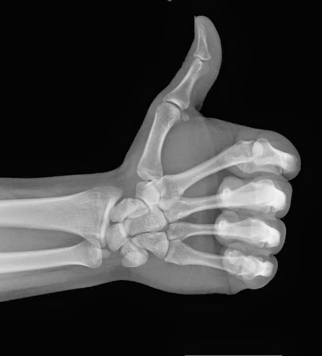
The advent of medical imaging dates all the way back to 1895, following the discovery of X-rays by the German physicist, Wilhelm Conrad Roentgen. The first X-ray picture was then produced, detailing the skeletal composition of his wife’s left hand. However, the actual quality of this imaging process was still very primitive, only allowing for the visualization of bones or foreign objects.
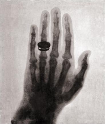 |
| Much to Dr. Roentgen’s pleasure, Mrs. Roentgen had not discarded her wedding ring |
- Ultrasound – Uses sound waves that are able to penetrate cellular tissue. Once they reflect off the body’s internal organs, the vibrations generate an electrical pulse which can then be reconstructed into an image.
- PET-CT Scan – Positron emission tomography (PET) uses compounds that emit positrons when they decay rather than gamma rays. It is now combined with a computed tomography (CT) device to generate a high-resolution image displaying sectioned layers of the scanned area.
- MRI – A Magnetic Resonance Imaging scanner runs a strong magnetic field through the body, aligning hydrogen protons. As the protons return to their original position in the atom, they generate radio waves, which are then picked up by the scanner and used to create an image based on signal strength.

Fast-forward to present day and over 70 million CT scans, 30 million MRI scans and 2 billion X-rays have been performed worldwide! The field of medical imaging is still growing by the day, with ongoing research leading to new developments.
Thanks for reading,