Research Opportunity Program: Cost Effectiveness and Imaging Carotid Stenosis

ROP Research Forum March 5th, 2015 – Sylvia Urbanik

Sylvia is a second year student studying cell and molecular biology. She is currently finishing up her Research Opportunity Program that spanned the fall and winter semesters, and also recently represented us at the March 5th ROP research forum along with her partner and predecessors.

 
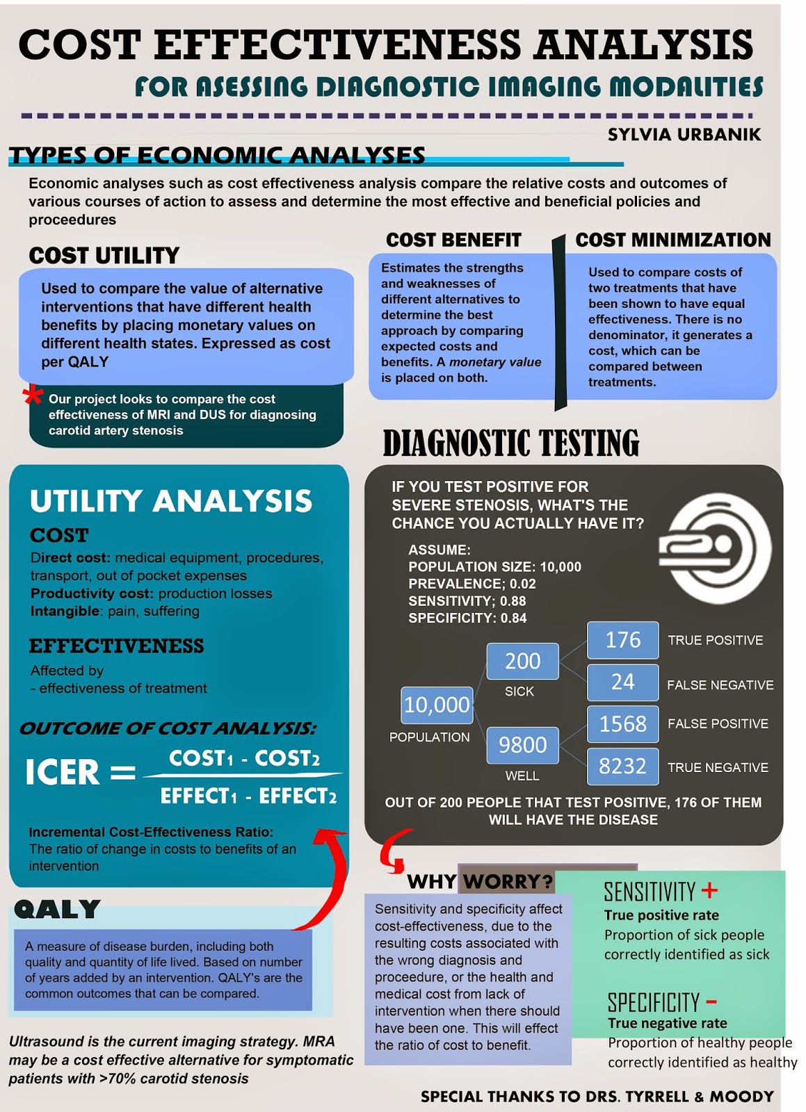
Her project dealt with cost-effectiveness analysis techniques for diagnostic imaging modalities, with a specific focus on carotid artery stenosis. She explored the different types of analyses used in assessing cost effectiveness. She examined how factors such as sensitivity and specificity of diagnostic tests (imaging modalities in this case) can affect the analysis, and conducted literature searches in order to find these variables in order to incorporate them into a cost effectiveness model.​

Well done, Sylvia!

Stay tuned, next Kevin Chen will be modeling with TreeAge…

See you in the blogosphere,

Pascal Tyrrell

Research Opportunity Program: Imaging Carotid Stenosis

ROP Research Forum March 5th, 2015 – Helena Lan

Helena is a second-year student pursuing a specialist in Pharmacology and Toxicology at the University of Toronto. Participating in ROP299 this past summer (see here) has opened many doors for her. She is currently assisting Pascal on a systematic review on research methodology and biostatistics in medical imaging, working as a research assistant for a medical education study, participating in the research abroad program at Karolinska Institutet this summer… and recently represented us at the March 5th, 2015 ROP research forum!

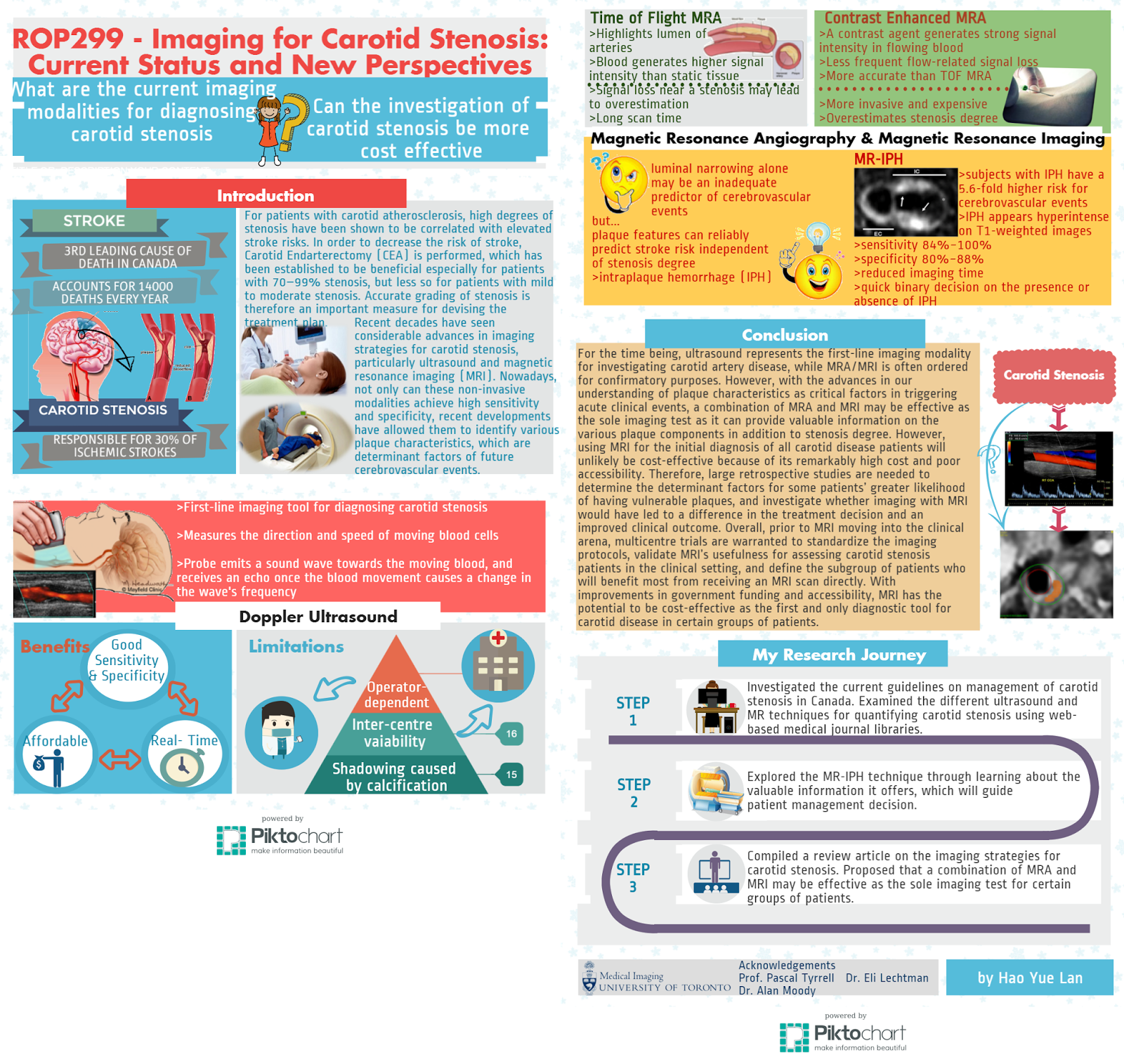
Helena examined the technical aspects and information provided by two major imaging modalities, MRI and ultrasound, for diagnosing carotid stenosis. She suggests that MRI holds great promise to serve as a cost effective test for carotid stenosis as well as a tool for assessing vessel health and plaque composition that would provide important information for patient management decisions.


Stay tuned, next Sylvia Urbanik will be talking about cost effectiveness…

See you in the blogosphere,

Pascal Tyrrell

Research Opportunity Program: What Is Carotid Stenosis and How Are We Treating Affected Patients?

ROP Research Forum March 5th, 2015 – Alana Man

Alana Man is a second year University of Toronto student pursuing a specialist in Bioinformatics and Computational Biology and a major in Immunology. She was a Research Opportunity student last summer with MiVIP (see here) and recently represented us at the March 5th, 2015 ROP research forum.

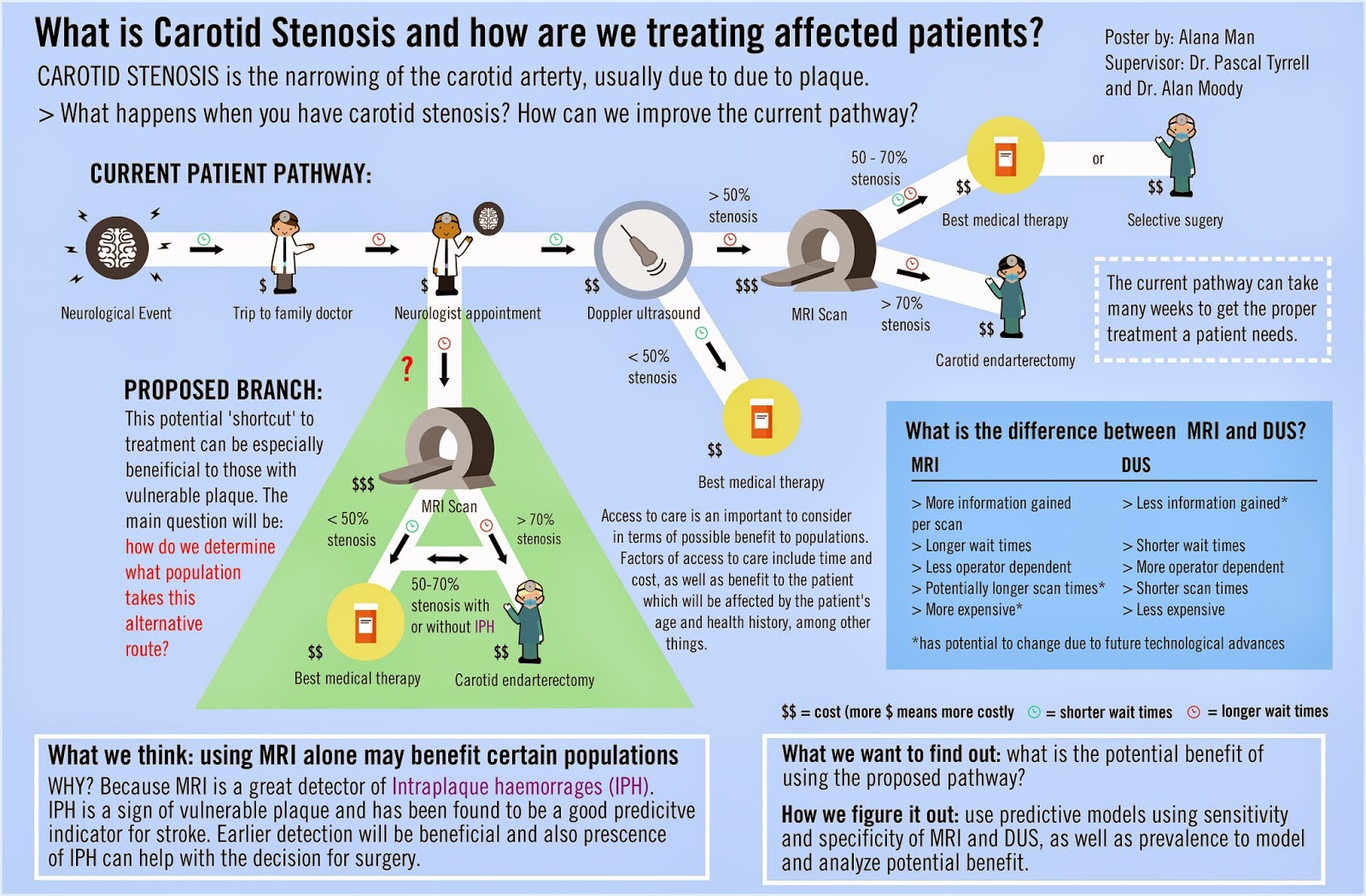
Alana’s project focused on care for patients with carotid stenosis in Toronto, Ontario, Canada. She looked at the different factors contributing to access to diagnostic testing such as procedure costs, wait times, and the differences between available imaging modalities and explores whether MRI could be the diagnostic test of choice over DUS for people at increased risk of carotid stenosis.

Well done, Alana!


Stay tuned, next Helena Lan will be comparing MRI and DUS…


See you in the blogosphere,


Pascal Tyrrell

The Adventures of Alana the Newbie Researcher

Timeline of my ROP experience

Stroke. Atherosclerosis. Carotid stenosis. MRI. Endarterectomy. To some people, these words are, well, just words. But for others, these few phrases could change the course of their lives. During my time in the Research Opportunity Program at the Department of Medical Imaging, I was able to learn all about carotid disease and the current status of carotid imaging modalities. As an added bonus, I did quite a bit of research on the impact that stenosis has on patient care and how new and exciting advances in medical imaging could affect it.


Over the course of four months, I went from being a huge research newbie to becoming an experienced research … trainee. I was introduced to the world of journal article writing, starting from doing elementary literary research all the way to drafting and writing a review article, which was completed entirely from the comfort of my beloved computer. With the help of Doctor Eli Lechtman and my ROP partner Helena Lan (if you haven’t seen her post on her experience you should check it out), we were able to put together 2 review articles that complement one another and will hopefully pose as a good foundation for future research.


Now, you must be thinking, “wow, this sounds like an extremely exciting summer; how could it possibly be better?” Well, my dear readers, it gets even better. One of the greatest things about being a part of this department is the community. The people here are all friendly, wonderful, and welcoming. From having an outing at Centre Island to letting us operate an uItrasound machine (thanks Modele!), I really felt like I was a part of “the gang.” What’s more, it was nice to have someone remember my name when I arrived in the morning, especially when I’m used to being my nameless self in the vast sea of students of our first year lectures (Convocation Hall, anyone?).


When I first sat down to write this post, I couldn’t decide what would be the best way to describe my time doing my ROP. And that’s when it hit me: it was an adventure! I may not have explored unknown lands or survived any near-death experiences, but there was not one day that went by where I wasn’t learning something new. Each day was a new experience, an opportunity to understand something I probably haven’t even heard of before. It still surprises me right now, as I’m writing this post, how much I have learned throughout this program, and the fact that I was able to experience something like this. I would have never thought I could write a review article and do the things I’ve done in only my first year of university. All in all, this was a great experience, and I encourage everyone to try out something like this!


Thanks for reading this, y’all! And if you just scrolled to the bottom of this post and thought “tl;dr,” you can still check out my infographic timeline (there’s pretty pictures)! Also I’d like to give a shoutout to my awesome Professor, Dr. Pascal Tyrrell (check out his latest post), as well as all the lovely members of the Medical Imaging department at U of T for making my ROP experience the way it turned out to be.


Remember to make every day an adventure,


Alana Man 🙂

Helena Lan: Summer 2014 ROP

Helena Lan Summer 2014 ROP

What is research like? If you had asked me this
question several months ago, I would have answered, “You wear a lab coat and
goggles while mixing chemicals or observing organisms. Hopefully something
interesting will happen, so that you get to publish your findings!” Well, after
participating in the Research Opportunity Program (ROP) at the University of
Toronto, I discovered that medical imaging research is more than just
pipetting, and is all the more exciting!

 
So what kind of research is conducted in the medical imaging world? For my ROP, the objective of my project was to evaluate the roles of the non-invasive imaging modalities for diagnosing carotid stenosis. Hence, I engaged in online literature research of the various imaging techniques for assessing this disease. In this process, I also learned to use Zotero to manage all my references, which provides an easy way to generate a bibliography (when the software doesn’t crash every time you open it). After gathering all the pertinent information, I then put together a review article suggesting how a change in the current imaging approach could potentially improve clinical outcome. Who knew a report could be compiled without doing the lab grunt work?     
 
Wait, so this is all a radiologist does? Sitting in front of a computer and typing all day? Of course not! During our time at Sunnybrook Hospital, we got the chance to chat with a radiologist and discovered that she could decide whether patients should be released after taking a look at their diagnostic images. Pretty powerful, eh? That’s not all. We also found out how radiologists identified any abnormalities in patients, as we had the opportunity to work with the VesselMass software which allowed for the delineation of the lumen and vessel wall of arteries on MRI images. Oh, and did I mention we observed an MRI and an ultrasound examination of the carotid arteries, and even got to perform an ultrasound scan ourselves. Super cool!
 
Still craving for more of my ROP experience? Check out my timeline infographic! You will find all the things I learned and all the fun I had there. Last but not least, I’d like to shout out a big THANK YOU to Prof. Pascal Tyrrell and Dr. Eli Lechtman, who guided us every step of the
way. Also, I’m very grateful to Dr. Alan Moody for including us in his research program at Sunnybrook, as well as other members of the VBIRG group who gave us the chance to
participate in various activities. My summer would not have been this fun and meaningful without all of your help!
 
Have fun researching,


Helena Lan
 

A YSP Student Perspective: MRI and Carotid Artery Disease

Hershel Stark, MED YSP 2014 Student

Throughout the month of July, I participated in a research program with the Division of Teaching Laboratories within the Faculty of Medicine at the University of Toronto. I was assigned to work with Prof. Pascal
Tyrrell and the Department of Medical Imaging, and spent the majority of my time with the Vascular Biology Imaging Research Group (VBIRG) at Sunnybrook Research Institute. I would like to discuss my experiences, what I gained from the program, and how I can take those skills with me into the future.


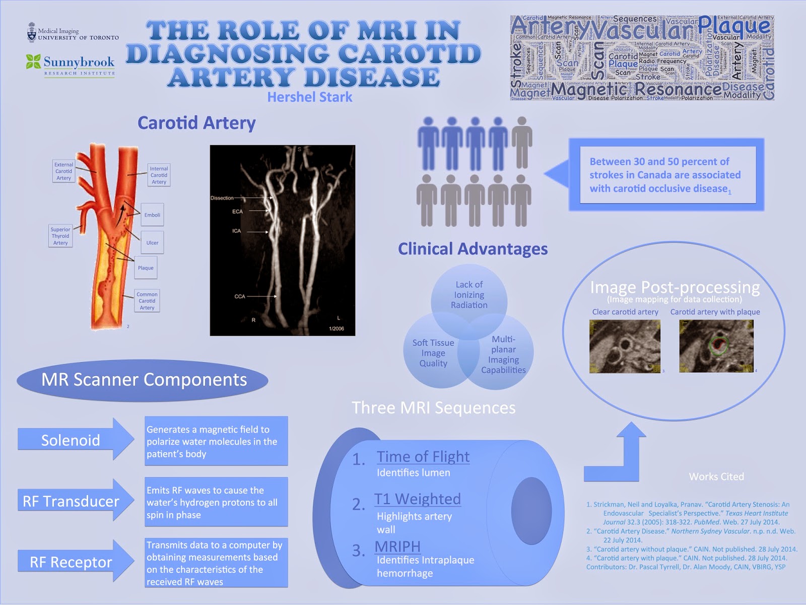
Essentially, the program was composed of presentations and shadowing opportunities in which I was introduced to various imaging modalities used in both the clinical and research fields. I primarily studied MR imaging, but was nevertheless exposed to other modalities including ultrasound and CT.  Towards the end of the program, I had two principal objectives: to present my experiences to the VBIRG group and to design an infographic for displaying. Below is a copy of my infographic:

Notwithstanding the abundance of knowledge I gained from studying the subject content, I acquired a variety of essential research skills by partaking in the program. Shadowing proficient researchers as they collected
and analyzed data provided me with a thorough insight of a researcher’s methods and techniques. The researchers that I worked with appropriately explained their individual roles on the research team, which led to my understanding of the significance of collaboration in scientific and medical research.
One last aspect of the program that I would like to address is the daily workshops that were conducted by two instructors from the Division of Teaching Laboratories, Jastaran Singh and Jabir Mohamed. Each of these brief workshops focused on an important general topic relevant to research in general, ranging from discussing common scientific practices to elaborating on literary research. I believe that the combination of skills and knowledge that I obtained from all elements of the program will be useful in my potential
research career in university.
 Lastly, I would like to take this opportunity to formally thank all of those that contributed to making the program a truly enjoyable and intellectually stimulating experience. I would like to extend my gratitude to
Dr. Alan Moody and the members of the VBIRG group for allowing me to shadow their research projects, as well as to Prof. Pascal Tyrrell and the Department of Medical Imaging at U of T for constructing the program and offering much assistance in the formation of my infographic. Finally, I’d like to thank Dr. Chris Perumalla and the Division of Teaching Laboratories in the Faculty of Medicine at U of T for formulating the research module of the Youth Summer Program, and Jastaran Singh and Jabir Mohamed for providing guidance as instructors throughout the program.
Best of luck in all of your future endeavours,
Hershel Stark
A YSP Student Perspective: Ultrasound – Not Just for the Unborn Child

Angela Lo, MED YSP 2014 Student
When you think of ultrasound, what’s the first thing you think of? Babies. All that fun stuff. Well, it turns out that ultrasound can be used not only for clinical testing, but also for research purposes. For
example, it can be used as a diagnostic tool to survey images of the body or also used as a device that monitors health conditions in research studies.
During the past three weeks, I (Angela!) have had the opportunity to participate in a research module in the medical imaging department at The University of Toronto. Through this program, I have been exposed to various imaging modalities including both MRI machines and CT scanners, but one of the modalities that interested me most was the ultrasound machine. Why? Because of its noninvasive procedures and its ability to make both 2D and 3D images in real time while still being a fraction of  he cost of an MRI.
Amazing.
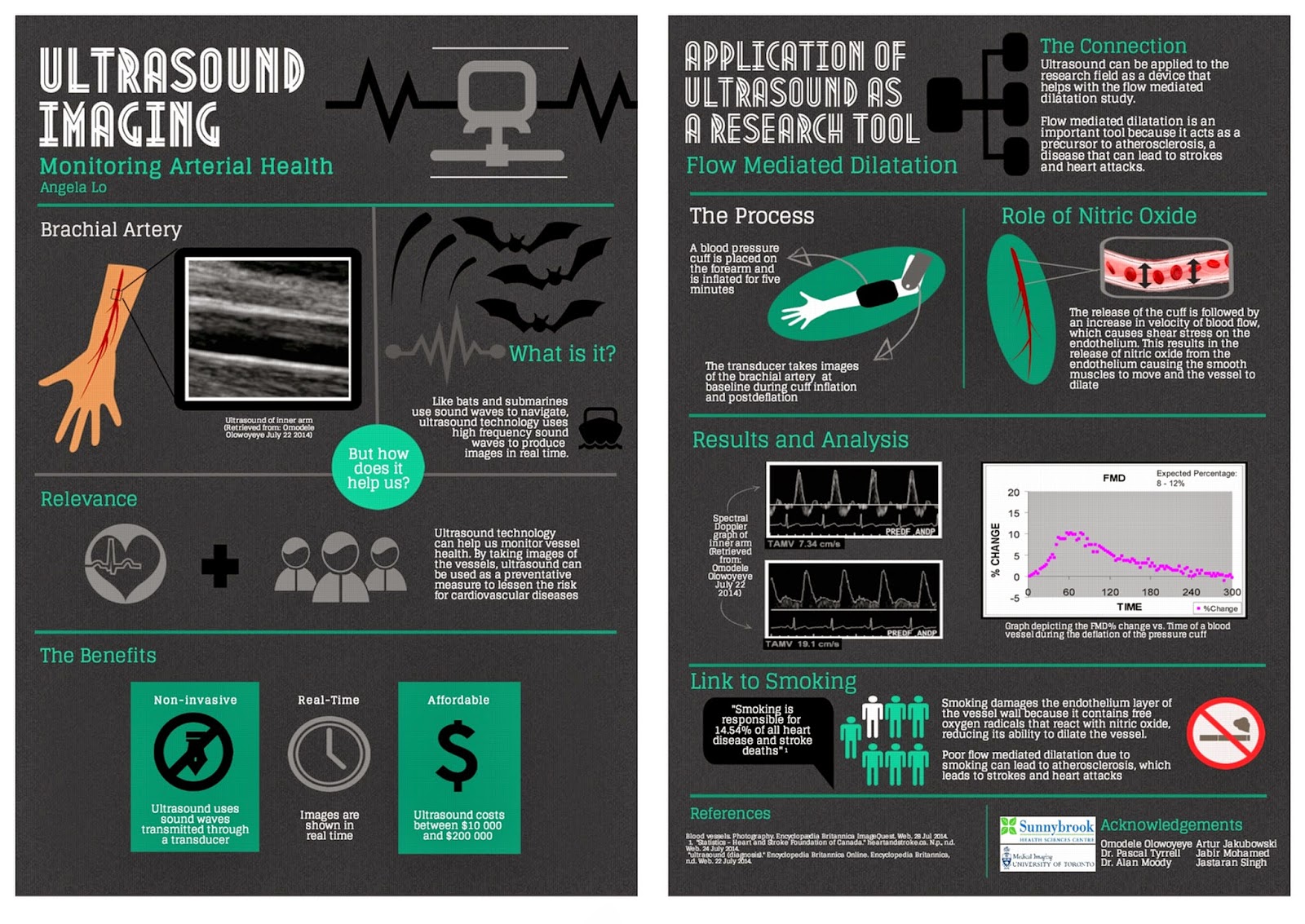
During my time in the program, I was also able to observe the various uses of the ultrasound machine and how it can be used as a research tool in the flow mediated dilatation study. Blood vessel health can be studied by having an ultrasound take images of the brachial artery to measure blood velocity and the percent change in FMD. By knowing the percent change, researchers can monitor arterial health and use it as a preventative measure.
Overall I had an amazing experience learning about all he imaging modalities and the great benefits and potential each one holds.
Happy reading,
Angela Lo

Michener Institute Series: Princess Margaret Hospital, Toronto, Ontario


As first year Radiation Thearpy students here a The Michener Institute, we are currently in our 4th week of clinical placements! As promised, here’s a little update about the experiences Jennifer and Ori are going through at Princess Margaret Hospital.

Jennifer: I’ve been placed in Unit 10 which specializes in treating patients with Genitourinary, Gynae and Lower Gastrointestinal cancer.

Ori: I’m on Unit 14 and we treat breast cancer and palliative cancers.

We are proud to say that we are enjoying our experience here. Our duty as students in training is to follow the radiation therapist and learn what they do. The job of a therapist is to treat cancer using a machine called Linear Accelerator (Linac) to deliver ionizing radiation. Patients will typically come once a day for the next couple of weeks, so we see the same patients every day and therefore really get to know our patients well. There is a fair amount of patient interaction, which is one of our favorite parts of the job. Along with patient interactions, we also get to use the equipment, which mainly includes operating the Linac machine (the machine that delivers the radiation) and taking X-rays or CT scans to make sure the patient is in the right position. Every day is a new experience and we are constantly learning new skills. We get a better insight of the patient’s perspective during the entire span of their radiation treatment. For example most patients in unit 10 are required to have a full bladder and empty rectum. Having to hold their pee can be quite difficult for some patients, especially when there are delays, which pushes Unit 10 to be a very fast paced environment. Overall our first 4 weeks of clinical has been an exceptionally valuable experience and we’re looking forward to our next 4 weeks!


Until next time!

Jennifer and Ori


Michener Institute Series: Clinical Placement Site – Kingston Ontario


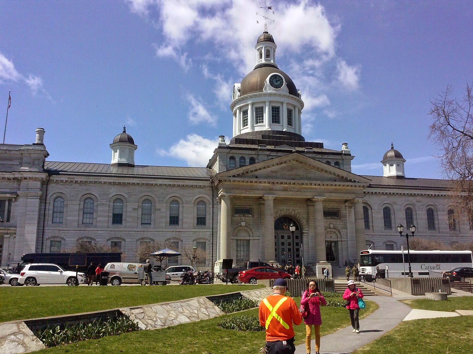
(Kingston City Hall)
It has been a month since the start of summer clinical placement, and I am currently
completing my placement in Kingston General Hospital (KGH) here at Kingston, Ontario.  Kingston is a nice beautiful town located at the north side of the entrance of outflow of St Lawrence River from Lake Ontario; it was the
first capital of Canada when Canada was still a province of British colony.
 
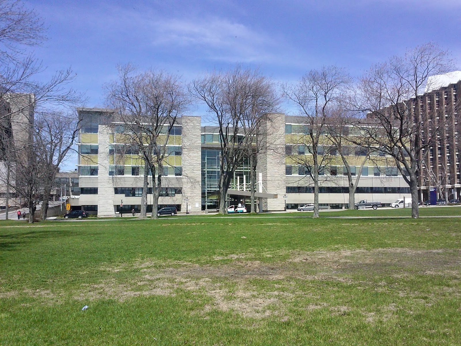
KGH host one of the most eastern cancer center in Ontario and it has a beautiful view because it is situated by the side of Lake Ontario, its front entrance open to the water. It is a perfect place for lunch and enjoys the sun during summer time.


      (KGH cancer centre front entrance)    
               
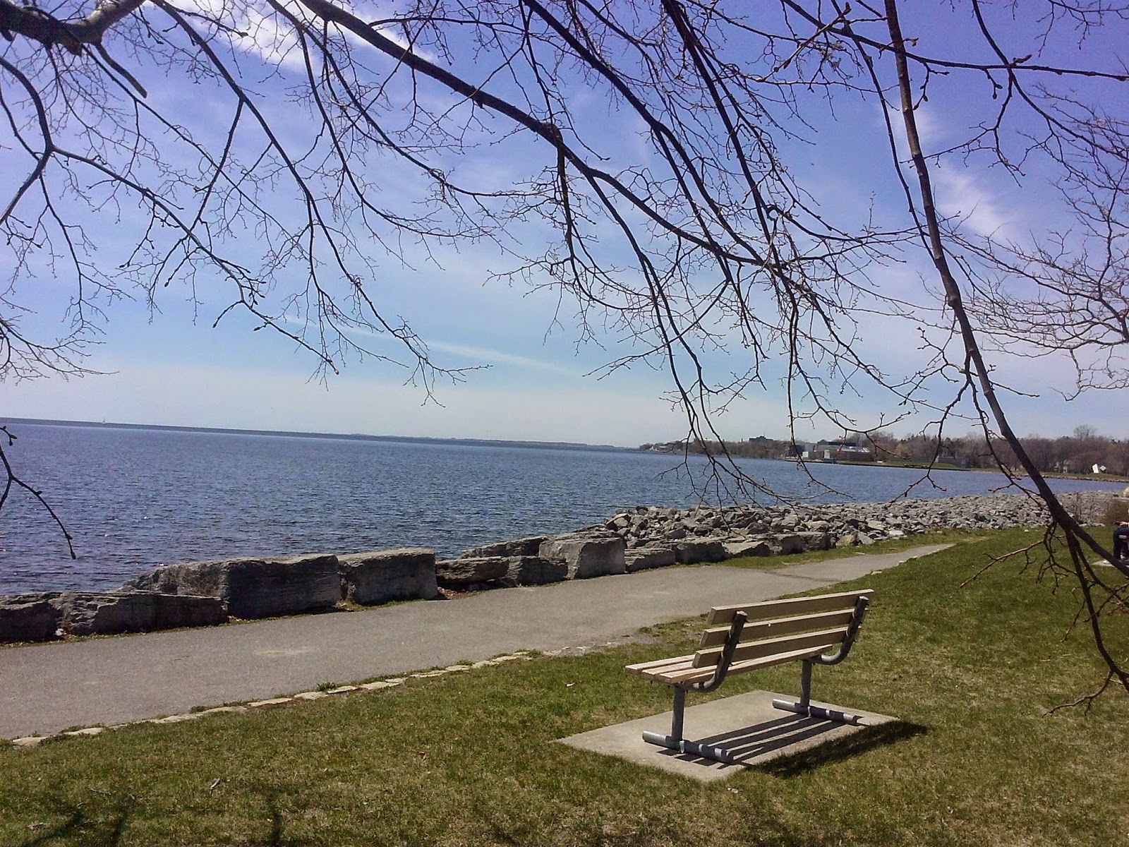
          (MacDonald
Park by water, in front of cancer centre)


The past month was phenomenal, words cannot fully describe the knowledge and experience we gain from clinical practice. The transition from purely academic to hands on
practice is eye-opening and a bit hectic; because each patient is unique and no knowledge from books can prepare you how to interact with all patients.  It is interesting to learn from the therapists, the way they educate patients on their first day of treatment, the type of approach to each patient base on the assessment they do during the conversation with them. It’s amazing how much compassion the therapists have for patients and how much they care for them.
 
 
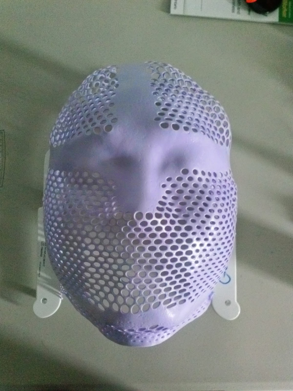
During the first two weeks in CT simulation unit, I made my first mask and had my own mask made for treatment to head and neck regions. The mask is made of pliable plastics. They come in as a sheet of plastic in a frame, and are put into a warm/hot water bath for 2-4 minutes to makes it pliable, after the mask is taken out of the warm water bath there is a 30-60 seconds window before it hardens. The therapist takes out the mask, tower dry it as much as possible and covers it on patient’s head as fast as possible.  The therapists are very efficient at their job, but what is amazing are the patients going through the process; imagine a warm and moist piece of plastic cover you face, harden in an instant and lock your head into position, and afterword you cannot move for 5-10 minutes for CT scan. I never had thought of the discomfort till I experience it myself.


(My 1st  mask, can kinda see my face print)
 
So far the experience here is amazing, and hopefully the coming June will be equally fantastic as well.
 
Till next time.


Gordon

Acting 101 – Radiation Therapy Role Play Exercise

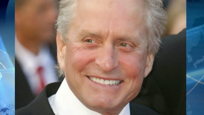
Michael Douglas: actor extraordinaire!

How is acting and radiation therapy related? Here at the Michener Institute, there are actual actors coming in to perform as our patients during patient care simulation and practical assessments. This is very helpful and fun at the same time as we get some experience with “patients” and if we make any mistakes, all can be corrected before going into the real world. This can spare us some embarrassment – the first time I talked with a patient actor, I could not think of what to say so “I am drawing a blank” just slipped out of my mouth! At the end of the debrief, the actor told me I could have just pretended to know by acting like The Thinker! Looking sophisticated and deep in thought.

Beside the patient actors coming in, we also do role play in patient care labs – free acting lessons! Just the past Tuesday, we had a role play class for scenarios in patients with special needs. Some students are just natural actors/actresses, sometimes I wonder why they are not in acting. The class was very fun and educational and allowed us radiation therapy students to learn how the patients will react and how we can respond. Anyway, I wish I had video to show you how fun it was. If you are interested, you should apply to Michener next year and experience it yourself…

Until next time,

Gordon