My name is Lee Radigan and I am a non-degree student pursuing admittance to the Biostatistics Master’s program at the Dalla Lana School of Public Health. After returning for my 6th year studying statistics at The University of Toronto, I thought that this was a perfect time to reflect on my progress.
Since September, I have been working under Dr. Pascal Tyrrell’s guidance on a project aimed at helping the Department of Medical Imaging report agreement in their research. To do this, I created a flow chart to help guide the reader towards the proper method of agreement. Along with this, I conducted a simulation looking at a specific question pertaining to the Department.
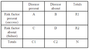
Initially, I was tasked with combing through various papers on the theory of agreement and making sense of all the different published work that was out there. There are many different approaches and different ways of looking at reporting agreement, so it was quite difficult to figure out when and where to properly use every single approach. After reading and re-reading each paper, as well as consulting the MiData team, I started to develop a thorough understanding of what agreement was, why it is important to report it, and how to go about reporting it appropriately.
Next, a flow chart was required to summarize what I had learned from the literature. This was not an easy task, because it forced me to dig really deep and make sure that every node in my flow chart was well thought out and appropriate. After many iterations and adjustments, I created a detailed chart that walks the reader from their initial research question up to the required agreement statistic.
My final task was to conduct a simulation that would test the question: Can a group of less experienced student raters be as accurate as a smaller set of more experience expert raters? And if so, how many students? And under what conditions? This was a very fun and informative task for me as I was able to conduct my first simulation. During this experience, my biggest difficulty was justifying my choices of parameters within the simulation. When conducting a simulation you have freedom to choose how it is going to work, but you must be careful to be able to back up each and every parameter choice. The simulation ended up showing that: the larger the disparity between the rating errors of the student and expert raters, the more students it takes to match the accuracy of the experts, confirming my intuition.
There are many things that I wish to expand on with respect to my project in future. I want to create a user friendly app that will be even easier and more compact than my flow chart. Additionally, I want to try to get my paper published. To do this I will need to look further into my simulation and consider a more broad range of student/expert scenarios that likely will occur in practice. I will also need to further refine my definitions and understanding of each concept of agreement.
This year has truly been the best of my life and I can largely attribute that to Dr. Pascal and the MiData team. I look forward to contributing to Medical Imaging research and to many more learning experiences.
Time to enjoy the summer as I embark on yet another exciting experience as a student Statistical Analyst at the CAMH Nicotine Dependence Clinic as a summer placement!
Lee Radigan