Stanley Hua in ROP299: Joining the Tyrrell Lab during a Pandemic

My name is Stanley Hua, and I’ve just finished my 2nd year in the bioinformatics program. I have also just wrapped up my ROP299 with Professor Pascal. Though I have yet to see his face outside of my monitor screen, I cannot begin to express how grateful I am for the time I’ve been spending at the lab. I remember very clearly the first question he asked me during my interview: “Why should I even listen to you?” Frankly, I had no good answer, and I thought that the meeting didn’t go as well as I’d hoped. Nevertheless, he gave me a chance, and everything began from there.

Initially, I got involved with quality assessment of Multiple Sclerosis and Vasculitis 3D MRI images along with Jason and Amar. Here, I got introduced to the many things Dmitrii can complain about taking brain MRI images. Things such as scanner bias, artifacts, types of imaging modalities and prevalence of disease play a role in how we can leverage these medical images in training predictive models.

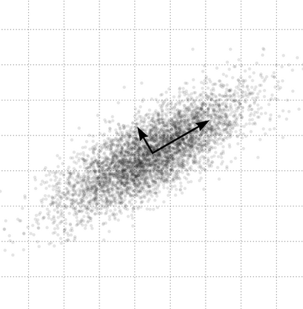
My actual ROP, however, revolved around a niche topic in Mauro and Amar’s project. Their project sought to understand the effect of dataset heterogeneity in training Convolutional Neural Networks (CNN) by cluster analysis of CNN-extracted image features. Upon extraction of image features using a trained CNN, we end up with high-dimensional vectors representing each image. As a preprocessing step, the dimensionality of the features is reduced by transformation via Principal Component Analysis, then selecting a number of principal components (PC) to keep (e.g. 10 PCs). The question must then be asked: How many principal components should we use in their methodology? Though it’s a very simple question, I took way too many detours to answer this question. I looked at the difference between standardization vs. no standardization before PCA, nonlinear dimensionality reduction techniques (e.g. autoencoder) and comparisons of neural network image representation (via SVCCA) among other things. Finally, I proposed an equally simple method for determining the number of PCs to use in this context, which is the minimum number of PCs that gives the most frequent resulting value (from the original methodology).

Regardless of the difficulty of the question I sought to answer, I learned more about practices in research, and I even learned about how research and industry intermingle. I only have Professor Pascal to thank for always explaining things in a way that a dummy such as me would understand. Moreover, Professor Pascal always focused on impact; is what you’re doing meaningful and what are its applications?

 I believe that the time I spent with the lab has been worthwhile. It was also here that I discovered that my passion to pursue data science trumps my passion to pursue medical school (big thanks to Jason, Indranil and Amar for breaking my dreams). Currently, I look towards a future, where I can drive impact with data; maybe even in the field of personalized medicine or computational biology. Whoever is reading this, feel free to reach out! Hopefully, I’ll be the next Elon Musk by then…

Transiently signing out,

Stanley Bryan Z. Hua

ROP299 2017-2018: A Medical Imaging Journey from a Humanities Perspective

My name is Samantha Santoro, and I am completing my second year in the English and Biology majors at the University of Toronto, St. George. A rather unconventional combination, when reviewing past students of Dr. Tyrrell’s lab. I was a 2017-2018 Research Opportunity Program (ROP) student in Dr. Pascal Tyrrell’s lab, and my work chiefly consisted of evaluating the internal vessel wall volumes of carotid arteries in a particular cohort of patients provided by the ongoing prospective CAIN study. My ROP was in the field of Medical Imaging. I am the co-president of the student club known as Watsi, with the main chapter based in San Francisco. I am also a special contributor to the Rare Disease Review, along with volunteering at an amalgamation of charity walks and fundraisers.

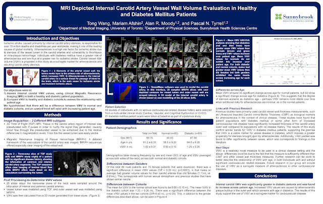
My ROP project was a turbulent experience – although that word is typically associated with a negative connotation, I regard my ROP299Y1 as one of the most humbling, interesting, and educative experiences that I have had thus far – most definitely not negative. However, to say everything went smoothly would be discrediting the lessons I learned from when things were not idyllic and smooth. My project, as aforementioned, statistically analyzed data provided by patients part of the CAIN study (an analysis that could not have existed without Dr. Tyrrell’s generous and unwavering support). My study determined that patients who were found to have IPH, or what is known as intraplaque hemorrhage, when I analyzed their MRIs, were also found to have increased vessel wall volume. This conclusion is incredibly significant, as IPH is a surrogate marker for atherosclerosis and could potentially be an indicator for patients at risk of future cerebrovascular events (namely, ischemic stroke). As strokes are currently the number three killer in the U.S and Canada alone, and heart disease number one, having a potential indicator for patients at risk of stroke would greatly benefit clinicians in their practice, as well as patients themselves.

As aforementioned, studies similar to my own are currently underway by the Canadian Atherosclerosis Imaging Network, furthering the important research in this field. The VBIRG (Vascular Biology Imaging Research Group) was the lab in which I primarily worked throughout the course of my ROP, at Sunnybrook Hospital. Moreover, I also worked on systematic reviews and reports outside of the focus of my project, in the fields of medical ethics and AI in the radiology workplace – both of which were opportunities provided to me by Dr. Tyrrell, and both of which were incredibly valuable experiences, allowing for me to broaden my knowledge of certain areas of medicine and science that are developing and expanding.

Although my project was littered with its own respective difficulties – a substantial number of drafts throughout each step of the program (more than I had ever made, even being an English student); a reluctant, but later fulfilling, acquaintanceship with the post-processing software VesselMass; and several late nights learning about the field of statistics – it is in light of these difficulties, and at present having overcome them throughout my ROP, that I remember Dr. Paul Kalanithi’s words in his memoir When Breath Becomes Air: “It occurred to me that my relationship with statistics changed as soon as I became one”. He, too, had studied Biology and English. I may not have played a lead role in the statistics I had been working with, but I can now say that understanding what they meant and how they were formulated has generated a deep respect in me for the field of statistics.

My poster was on display at the 2018 Research Opportunity Undergraduate Fair. Special thanks to Mariam Afshin, my supervisor at Sunnybrook Hospital; Bowen Zhang, for answering each question I had while at Sunnybrook; John, and the rest of the lab team; and Dr. Pascal Tyrrell, for answering my email last February and holding my interview on the same day as my Chemistry exam. Never before had I met such an – in a word – outstanding professor, and I dare say that I will never meet one like him throughout the rest of my academic journey.

Samantha Santoro

MRI, Statistics, Carotid Arteries, and 1000 Cups of Coffee with George Wang

GeorgeWang – ROP299Y 2016-17
I’m George. I have recently completed my 2nd year undergrad at the University of Toronto studying physiology and physics. In the fall-winter term of 2016-17 I had the privilege to work in Pascal’s group, looking into carotid artery MRI and using the volume of the carotid artery vessel wall as a marker for atherosclerosis. Having an acquired interest in medical imaging and a previous summer position working with PET, I saw this as an excellent opportunity to expand my knowledge of the field while having the chance to be exposed to clinical research methods. Above is my account of how the year went in a nutshell.
 
Have a look at my poster from the ROP Research Day below…


Wow! What a Busy Summer….

Jenny Joo – YSP 2016

Over 20 students in the lab this summer beavering away at some great projects. Last week my two Youth Summer Program (University of Toronto) students finished their three week stay with us. 


Jenny and Michelle both did fantastic work.


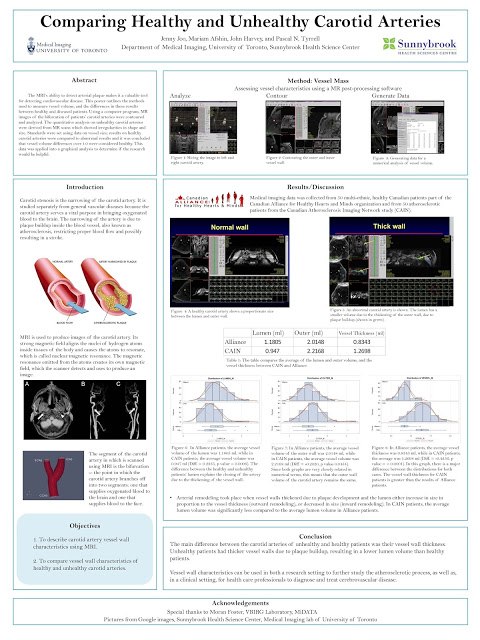
Today Jenny will show you her poster entitled:“Comparing Healthy and Unhealthy Carotid Arteries”


Jenny Joo is from Richmond Hill, Ontario, entering her senior year of high school. She plans on studying life science at the University
of Toronto in the future. She spent the last 3 weeks in U of T’s YSP Medical
Research program, where she was placed in two different medical imaging labs: The
MiDATA lab of U of T and the Vascular Biology Imaging Research lab at
Sunnybrook Hospital. 

Jenny chose to do research on the
MRI scans of the carotid artery because it focused on both research and
clinical aspects and had this to say about her experience with us: “It has been an enriching 3 weeks working with my PI, Pascal
Tyrrell, my mentors, John Harvey and Moran Foster, and the rest of the research
group.” 

Great work Jenny Joo!


Have a peek at her poster and…

… I’ll see you in the blogosphere.


Pascal Tyrrell

U of T Research Opportunity Program – Clare Sheen

Clare Sheen is an undergraduate student at the University of Toronto, in process of completing her Bachelor of Sciences in Genomics and Microbiology/Molecular Genetics. She was a 2015-6 Research Opportunity Program (ROP) student working on designing the Medical Image Network Enterprise (MiNE) interface for Dr. Pascal Tyrrell from U of T’s Department of Medical Imaging. She is currently a social director on the Life Science Student Network exec team and a volunteer at U of T’s Agrawal Lab where she helps with Drosophila experiments. She continues to seasonally work as a student camp teacher in the summer.

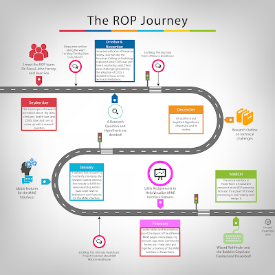
At the Research Opportunity Program (ROP) fair on March 3rd, U of T ROP students from different departments came together to share their research. A mock-up of the MiNE interface was presented in PowerPoint with the goal of increasing user engagement and encouraging the development of a medical imaging research community. Some features of the interface are presented below.

Another Reason Why a Brain Boo-Boo Is Bad.

Rostam Rashidkhani – YSP 2015


Rostam Rashidkhani is a grade 12 International Baccalaureate student at the Toronto French School and he was a Meds – Youth Summer Program student with me this summer.

Rostam is intrigued by the sciences and enjoys biology, chemistry, and physics in school. He has participated in a number of University of Toronto summer programs and is looking forward to University life!


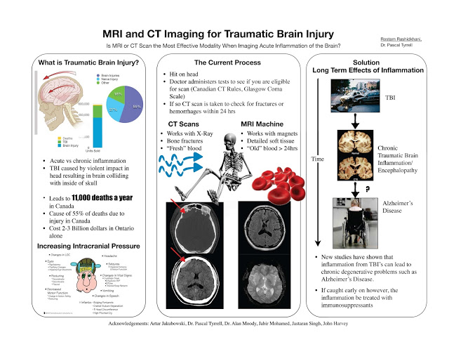
This summer Rostam looked at what causes brain problems after traumatic brain injury and how best to detect these changes with MRI. Recent brain imaging studies, including those in former professional football players, indicate that persistent brain inflammation after a single moderate head injury or repeated milder traumatic brain injury may be very common, may contribute to cognitive problems. More importantly, the chronic brain inflammation related to traumatic brain injury may be treatable. Looking for chronic traumatic brain inflammation with followup MR may be a way to reduce cognitive impairment.

Well done, Rostam!


Enjoy the read and…


… see you in the blogosphere,

Pascal Tyrrell

MiWord of the Day Is… UBO?!!!

I must mean UFO or Unidentified Flying Object? You remember the movie Close Encounters of the Thrid Kind? Spielberg’s massive hit in 1977 following his release of the original Jaws. Back in those days UFO sightings were often in the news (or tabloids anyway) and this movie hit the sweet spot. It even helped launch the toy “Simon” which as it turns out was very similar to the multicolored, note-playing alien saucers featured in the movie – coincidence?


So, what the heck is UBO? Well, as it turns out the human body exhibits a variety of anatomical details in the ever so important Magnetic Resonance Imaging (MRI) scan that we have all learned to love (see our series on MRI and Carotid Stenosis). The majority of patients have similar anatomical features on imaging but some fall outside these normative patterns. When radiologists come across findings that are difficult  to interpret they will often refer to them as “Unidentified Bright Objects”. The challenge, of course, is that the radiologist needs to decide whether to label the anatomy in the image an “UBO” – essentially an image artifact – or “disease”.


This is where the rubber meets the road. Interpretation of MRI scans is work done by people, and, as with all jobs, the quality of performance varies. Therefore, the accuracy of the MRI exam is heavily dependent on the quality of the radiologists who interpret them. It is for this reason that the training a radiologist receives is crucial to his/her success. In addition, there is an important relationship that exists between the radiologist and the primary care physician as they have to balance indications of abnormality in MRI scans with the information provided by other techniques such as the clinical exam. A successful diagnosis relies on a good team effort. 


Go Team!


Now for the fun part (see the rules here), using UBO in a sentence by the end of the day:

Serious: Went for my MRI today. Told me that the UBO on imaging was just an artifact. Nothing to worry about. Phew!

Less serious: Hey Bob, did you hear on the news the report of another UBO hovering over farmer John’s field last night?  Or was that UFO? I always get those two mixed up…


Listen to UB [4] 0’s Red Red Wine to decompress and…


…I’ll see you in the blogosphere.


Pascal Tyrrell

An “Egg-cellent” Journey to Investigate Carotid Artery Stenosis in Cambridge, Ontario…


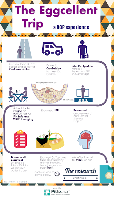
Last Wednesday, my ROP students Kiersten and Indranil, and UofT medical student Eli Lechtman had the opportunity to interview the legendary (see here) Dr. Kim Tysdale – a general practitioner in Cambridge, Ontario.


Why do you ask? Well each year 50,000 Canadians suffer from a stroke with 26% due to carotid artery disease. Carotid artery stenosis is the narrowing of the carotid arteries due to plaque buildup (see atherosclerosis). These plaques can then rupture and create blood clots that travel up to the brain. In turn, these blood clots then get stuck in the brain’s smaller blood vessels, causing a stroke. So plaque = bad and ruptured plaque = worse!


What if we had a clue as to which plaque may rupture? Well, in turns out that the presence of intraplaque hemorrhage (IPH) can help us predict just that! IPH is bleeding within the plaques, which causes them to become more vulnerable (see vulnerable plaque). There is a 6 times greater risk of stroke in people with IPH! And…. a new medical imaging technique called MRIPH imaging allows for visualization of IPH. MRIPH is similar to traditional MRI but highlights the artery walls and looks at the arterial plaques. 


While in Cambridge, Kiersten, Indranil, and Eli presented Dr. Tysdale with an overview of medical imaging techniques for the assessment of carotid artery stenosis with an emphasis on the new MRIPH technique. Informing physicians like Dr. Tysdale which of his patients are more at risk of having vulnerable plaques (by providing information on IPH) could result in patients receiving more appropriate and timely treatment – thereby reducing the number of strokes! 


BTW, we had Dr Tysdale at “hello” (not sure what I am referring to? See here).


So what up with the eggs? Kiersten, Indranil, and Eli also had the chance to tour Dr. Tysdale’s beautiful country house and meet his lovely wife (and nurse!), Eva, who sent everyone home with fresh chicken eggs! Awesome.


Thank you Dr Tysdale for being such a great sport!


Here is their infographic of the trip (great job gang!). Enjoy!

Kiersten Thomas and Indranil Balki – ROP Summer 2015


Stay tuned for more ROP adventures!


Pascal Tyrrell

MiWord of the Day Is… Magnet!

Who hasn’t thought of having Magneto’s powers? No? Maybe you should watch this Magneto trailer for a refresher. 


Ok, now that we all want to be Magneto (secretly at least) what is it that is so appealing with having the power of magnetism? Bill Nye the Science Guy explains it very well in this clip. Have a gander.


In a nutshell, magnetism is a physical phenomena that consists of a field of energy created by “magnets” that attracts or repels other objects. Magnets come in two major flavors: permanent magnets made of materials (such as iron) and electromagnets – the strongest and most widely used in medical imaging. 


Interestingly, it is the sum of the magnetic fields of individual electrons that is responsible for all the fun (see quantum mechanics). In the case of electromagnetism the electric current in a wire produces a magnetic field in the same direction of the current. In the case of a permanent magnet it is the magnetic fields of the naturally occurring electrically charged particles of the atoms that make up the material (iron for example) that are responsible. However, for there to exist a force strong enough to attract or repel another object all of its magnetic ions must have their magnetic fields aligned and contributing to the net magnetization. This is how you can magnetize a needle when stroking it in a uniform directional way with a permanent magnet.


Magnetism is to MRI what radiation is to X-rays. The strength of magnets is measured in gauss and Tesla units. There are 10,000 gauss to a Tesla and the earth’s magnetic field is one half of a gauss. Today most clinical MRIs use superconducting magnets whose strength range up to 4 Tesla! Experimental MRIs can run up to 10 Tesla. Now that is more Magneto’s speed.


The powerful magnets allow for better spacial resolution allowing for better sensitivity of the image. However, all this magnetic strength comes at a cost: the production of chemical shift artifacts – ghosts of things that are not really there. This is why we have radiologists to make sense of it all.


OK. Now you are asking what the heck. Magneto in the X-Men movie was able to rip out the iron from a human so why doesn’t an MRI? Great question. Iron found in the human body is mostly found as ferritin (a type of iron oxide) and is NOT magnetic. The iron in hemoglobin is also NOT magnetic. Bummer. So how does Magneto do it? Well either the movie is not scientifically correct (now that would be a shocker) or possibly he could be drawing on magnetite (another iron oxide) that is magnetic and has been found in trace amounts in the blood and brain. It is so little though that it does not cause any concern for MRI. Oh well, so much for Magneto…

Now for the fun part (see the rules here), using magnet in a sentence by the end of the day:

Serious: Hey Bob, did you know that early MRI machines used permanent magnets?

Less serious: Went for my MRI today. Told them I was worried the MRI would rip all the iron out of my blood like in X-Men. They didn’t even know who Magneto was. Whaaaat?!!

OK, listen to Magnetic by Traphik to decompress and I’ll see you in the blogosphere…

Pascal Tyrrell


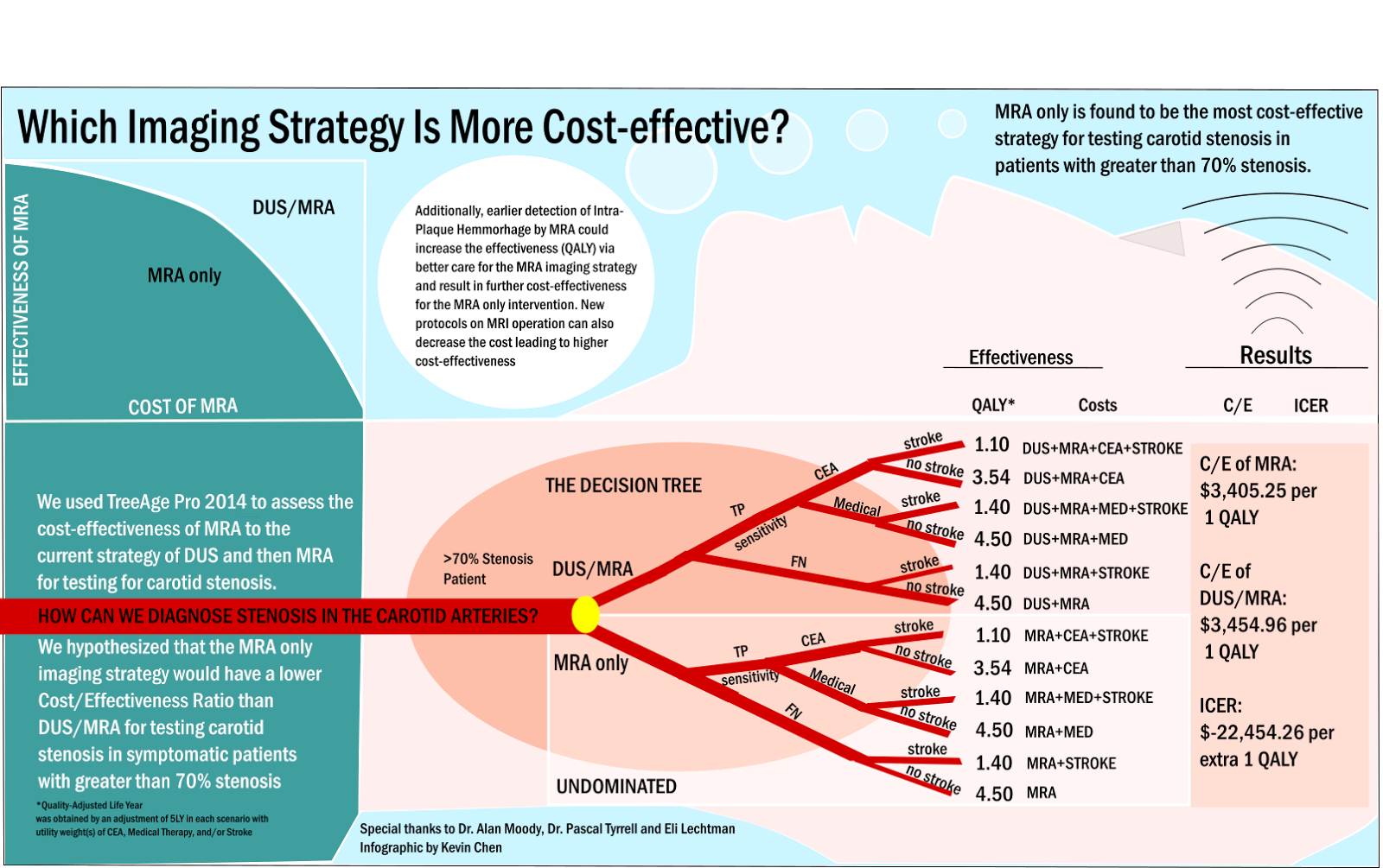
MRA Is More Cost-Effective than the Current Strategy for Testing > 70% Carotid Stenosis

ROP Research Forum March 5th, 2015 – Kevin Chen


Kevin Chen is a 2nd year research opportunity program student studying neuroscience and
physiology in the University of Toronto Life Sciences Program. His main goal is to get into
medical school and to enjoy the UofT experience as much as possible!

Kevin’s ROP project consisted
of cost effectiveness analysis modeling to explore whether MRA could be a
cost-effective measure in testing and treating patients with carotid stenosis.
Preliminary results showed that in a subset of the population at risk for
carotid stenosis (> 70 %), MRA was dominant over the current strategy of testing with
Doppler Ultrasound. By reducing MRA scan time and by tailoring MRA sequences we believe it possible to extend these findings to include a larger sub-population (> 50 %). More to follow…

Well done, Kevin!

See you in the blogosphere,

Pascal Tyrrell