MiWORD of the day is… Mop-top!

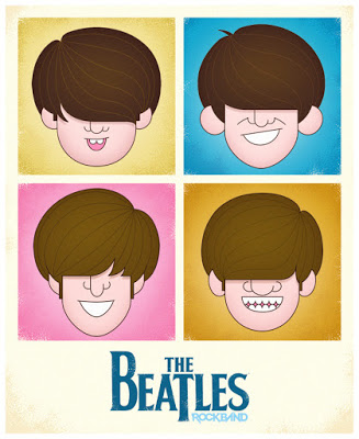
Ahhh, the mop-top! I sigh not because I miss the hairdo but because I miss my hair – all of it. In the mid-60s this hair style was made famous by The Beatles. Don’t know who they are (shame on you!) have a listen here for instruction.


Well the mop-top was made popular because the 4 guys who sported the hairdo were crazy successful musicians from England. Their recording company, Electrical Musical Industries (EMI), was also very happy and successful because of the overwhelming record sales (music was sold to listeners on vinyl records back then).


So, what does any of this have to do with medical imaging? Lots actually. The money generated by record sales enabled the EMI basic science researchers (another division of the company) to work in a prosperous cash-rich environment. One of those researchers was Sir Godfrey Hounsfield, an electrical and computer engineer. 


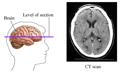
In 1967, he started his work on what would soon become the first CT scanner. By directing x-ray beams through the body at 1 degree angles, with a detector rotating in tandem on the other side, he was able to measure the attenuation of x-rays. These values were then analysed using a mathematical algorithm and a computer to yield a 2-D image of the interior of the body. The production of CT scanners by EMI started in the early 1970s and their monopoly ended by 1975 when companies like DISCO (not even kidding) and GE entered the arena.


Interestingly, in the 1960s Dr Allan Cormack of South Africa had also independently showed similar results to Housfield. In the end, Cormack was cited for his math analysis that led to the CT scan and Housfield for its practical development. They shared the Nobel prize in Physics and Medicine in 1979. Cool.

Now for the fun part (see the rules here), using mop-top in a sentence by the end of the day:

Serious: Who would have thought the success of the mop-top Fab Four would be instrumental in the development of the CT scanner?

Less serious: Hey Bob, I went for my head CT scan today and something weird happened. I went in bald and came out with a mop-top! Is that normal?…

Listen to With a Little Help from My Friends from The Beatles to decompress and…

…I’ll see you in the blogosphere.

Pascal Tyrrell


Some R&R with UofT YSP and Elizabeth Lehner…

Elizabeth Lehner – YSP 2015


Maybe not all rest and relaxation but certainly radiology and rheumatology! Here is a great example of why collaboration between disciplines is so important in medicine. Elizabeth recently graduated from Iroquois Ridge High School and will be a new University of Toronto student this fall. See her post below. 


Great job Elizabeth!!!

Many people are familiar with the word arthritis. This is probably because one in six Canadians aged 15 years and older report having arthritis. Rheumatoid Arthritis is a specific form of arthritis that unfortunately can lead to severe disability and joint replacement. 


Over the past several weeks, I participated in the 2015 YSP research program with the Division of Teaching Laboratories within the Faculty of Medicine at the University of Toronto and had the opportunity to look more closely at Rheumatoid Arthritis and ways to better diagnose this debilitating disease.


Under the supervision of Prof. Pascal Tyrrell and the Department of Medical Imaging at U of T, I was introduced to various imaging modalities including MRI machines, CT scanners and ultrasound machines. The work by Dr. Tyrrell was of particular interest given his studies on inflammation and the use of the various imaging modalities.


As part of this program I also participated in specific lab tasks including dissections and micropipetting and was exposed to clinical work such as suturing and operating an ultrasound machine. In addition, the program provided me with the opportunity to participate in daily workshops led by two instructors from the Division of Teaching Laboratories, Jastaran Singh and Jabir Mohamed. These workshops provided important overviews on a variety of topics relating to research that were very interesting.


The things I learned in this program provided me with a much better understanding of various research and medical issues that I think will be of use to me as I begin my studies at the University of Toronto this fall.
I would very much like to thank Prof. Pascal Tyrrell, Jastaran Singh and Jabir Mohamed for allowing me to be exposed to the various projects and for answering the many questions that I had during the program. Thank you!

 
Elizabeth Lehner

MiWord of the Day Is… Magnet!

Who hasn’t thought of having Magneto’s powers? No? Maybe you should watch this Magneto trailer for a refresher. 


Ok, now that we all want to be Magneto (secretly at least) what is it that is so appealing with having the power of magnetism? Bill Nye the Science Guy explains it very well in this clip. Have a gander.


In a nutshell, magnetism is a physical phenomena that consists of a field of energy created by “magnets” that attracts or repels other objects. Magnets come in two major flavors: permanent magnets made of materials (such as iron) and electromagnets – the strongest and most widely used in medical imaging. 


Interestingly, it is the sum of the magnetic fields of individual electrons that is responsible for all the fun (see quantum mechanics). In the case of electromagnetism the electric current in a wire produces a magnetic field in the same direction of the current. In the case of a permanent magnet it is the magnetic fields of the naturally occurring electrically charged particles of the atoms that make up the material (iron for example) that are responsible. However, for there to exist a force strong enough to attract or repel another object all of its magnetic ions must have their magnetic fields aligned and contributing to the net magnetization. This is how you can magnetize a needle when stroking it in a uniform directional way with a permanent magnet.


Magnetism is to MRI what radiation is to X-rays. The strength of magnets is measured in gauss and Tesla units. There are 10,000 gauss to a Tesla and the earth’s magnetic field is one half of a gauss. Today most clinical MRIs use superconducting magnets whose strength range up to 4 Tesla! Experimental MRIs can run up to 10 Tesla. Now that is more Magneto’s speed.


The powerful magnets allow for better spacial resolution allowing for better sensitivity of the image. However, all this magnetic strength comes at a cost: the production of chemical shift artifacts – ghosts of things that are not really there. This is why we have radiologists to make sense of it all.


OK. Now you are asking what the heck. Magneto in the X-Men movie was able to rip out the iron from a human so why doesn’t an MRI? Great question. Iron found in the human body is mostly found as ferritin (a type of iron oxide) and is NOT magnetic. The iron in hemoglobin is also NOT magnetic. Bummer. So how does Magneto do it? Well either the movie is not scientifically correct (now that would be a shocker) or possibly he could be drawing on magnetite (another iron oxide) that is magnetic and has been found in trace amounts in the blood and brain. It is so little though that it does not cause any concern for MRI. Oh well, so much for Magneto…

Now for the fun part (see the rules here), using magnet in a sentence by the end of the day:

Serious: Hey Bob, did you know that early MRI machines used permanent magnets?

Less serious: Went for my MRI today. Told them I was worried the MRI would rip all the iron out of my blood like in X-Men. They didn’t even know who Magneto was. Whaaaat?!!

OK, listen to Magnetic by Traphik to decompress and I’ll see you in the blogosphere…

Pascal Tyrrell


MiWord of the Day Is… Xeroradiography!

Who hasn’t done some creative photocopying at some point in their lives? I certainly do NOT condone this type of activity (very naughty) but would you believe me if I were to tell you that for a long while mammography made use of photocopy technology? Yes, I realize this sounds a little funny. Let me explain.


In the 1970s medicine made the association between heavy exposure to radiation for TB and thyroid treatments and the appearance of breast cancer three decades later. A reevaluation of the effects of radiation ensued and a call for ways to minimize exposure to ionizing radiation was made to the industry.


One of the first to answer that call was the radiologist John Wolfe from Detroit Receiving Hospital who in 1966 reported on the advantages of coupling photocopy technology with mammography. Xerox corporation jumped on the idea and developed a commercial unit in 1971 and “xeroradiography” was born! Basically, film from traditional x-ray imaging (yes back then they still used film!) was replaced with a selenium coated aluminum plate that was prepared for the exposure by being electrically charged. The result was that only a short burst of radiation (shorter exposure time means lower dose of radiation) was required to produce a very high quality image.


These xerox mammograms dominated the industry for over 20 years until new technology was developed more recently that provided even finer images with even less radiation. Cool.


Now for the fun part (see the rules here), using Xeroradiography in a sentence by the end of the day:


Serious: Hey Bob, did you know that mammograms produced using xeroradiography were blue? 


Less serious: My friend Jane was scheduled for a mammography. Having heard of xeroradiography reading the MiVIP blog she decided to DIY at her office. Problem was the print kept coming out black and white instead of blue from the Xerox machine…


OK, watch the Copy Cat trailer to decompress (or not?!!!) and I’ll see you in the blogosphere…


Pascal Tyrrell

MiWord of the Day Is… PET scan!


Peanuts. What a great story. The most popular and influential comic strip in history. Snoopy was my first stuffed animal growing up. He still lives with my parents. So what is a PET scan anyway? I don’t recall ever seeing the picture above in any of the Peanuts cartoon strips.


Positron emission tomography (PET) is somewhat of a special medical imaging modality in that it brings together two different technologies from different times. Let me explain. Back in the early 1930s, George Hevesy was a young Hungarian physicist who developed biologically safe and useful radioactive tracers that could be ingested or incorporated into the body in some way.  Physicians would then manually locate where these radioactive tracers had gone in the body by using a Geiger counter at first and then later using special cameras (Kuhl‘s photoscan) to produce a crude emission image. 


So, how do we get cool pictures like these ones?  Well we would have to wait another 25 years after the development of radioactive tracers by Hevesy for the start of construction of instruments able to not only detect these radioactive sources in the body but to produce tomographic pictures.


It won’t be until the mid 1970s that PET – as we know it today – would be born. Essentially, a patient receives a emissions scan (PET) and a CT (we talked about that here) or MRI (we talked about that here) scan at the same time. The two scans are then merged together thanks to highly specialized computers (see the pictures in the middle frames). Voila! PET. 


PET is both a medical and research tool. Most often used in clinical oncology (medical imaging of tumors and the search for metastases), it is also important in clinical diagnosis of certain diffuse brain diseases such Alzheimer’s disease and other types of dementia.


Relax your brain a little listening to Radioactive by Imagine Dragons and don’t forget the fun part (see the rules here), using PET scan in a sentence by the end of the day:

Serious: Hey Bob, did you know that much of the success of the PET scan is due to the development of the radiopharmaceutical FDG (deoxyglucose) that lead the way to the characterization of Parkinson’s and Huntington’s disease?

Less serious: I can’t believe they developed yet another PET scan. Wasn’t the CAT scan enough?

See you in the blogosphere,

Pascal Tyrrell


MiWord of the Day Is… Haptoglobin!


Just got back from the RSNA! Wow what a big conference – 56,000 people this year. McCormick place in Chicago, Illinois (where the conference is held) feels like an airport it is so big. 


Love Chicago. Great city. 


Of course, I had the pleasure of attending a bunch of great presentations and today I will introduce you to one of them. Tina Binesh Marvasti (say that 7 times fast!) presented on the topic of Haptoglobin. No, not Hobgoblin (not sure who that is? See here) or his infamous green predecessor (see here). 


So, what is Haptoglobin you ask? It is a serum protein that binds free hemoglobin – resulting from the breakdown of red blood cells – and functions to prevent loss of iron (contained in the heme group) through the kidneys and to protect tissues from the highly reactive heme groups. Essentially a housekeeping protein that helps to recycle hemoglobin as part of the red blood cell life cycle. Now what if your ability to clean-up free hemoglobin was impaired? Well, quite simply you would be putting at risk those sensitive tissues that come into contact with free hemoglobin. 


One important example of this is vessel walls affected by atheroma (AKA plaque). Sometimes these atheroma can bleed (called intraplaque hemorrhage or IPH) which worsens the whole situation. Typically, your body responds by sending the clean-up crew including the Hobgoblin (or haptoglobin, I always get these two confused). 


When people have the recessive genotype (Hp 2-2) of the Hp gene they produce less haptoglobin and therefore are at increased risk of damage from free hemoglobin (or more specifically the heme groups).


Tina and friends hypothesized the following: 


And she found that having the recessive Hp2-2 genotype was associated with a higher prevalence of IPH in a group of  80 patients (average age of 73 yrs). She also found that the IPH volume of Hp2-2 patients worsened over time.


So what is the take home? The Haptoglobin genotype is associated with IPH which is a biomarker of high risk vascular disease and could identify populations at higher risk of developing cardiovascular events.


Now for the fun part (see the rules here), using Haptoglobin in a sentence by the end of the day:

Serious: Hey Bob, did you know that a recessive haptoglobin genetype may contribute to an increased risk of cerebrovascular disease?

Less serious: My GP suggested that based on my recessive hobgoblin genotype I should consider a healthier lifestyle. Funny, I always figured Doc Ock to be the one to watch for…

OK, watch the Spider-man 2 trailer to decompress and I’ll see you in the blogosphere…


Pascal Tyrrell

MiWord of the Day Is… CAT scan!

A what scan? I am actually a cat guy myself. Not to say I don’t love dogs but if I had to make a choice…


I just finished reading a fantastic book by David Dosa entitled “Making Rounds With Oscar”. The premise of the book is a story about an extraordinary cat but the subject matter is very serious –  dementia and end-of-life care in the elderly. Have a gander.


So what the heck is a cat scan and what does it have to do with medical imaging? 


CT scans – also referred to as computerized axial tomography (CAT) – are special X-ray tests that produce cross-sectional images of the body using X-rays and a computer. CT was developed independently by a British engineer named Sir Godfrey Hounsfield and Dr. Alan Cormack and were jointly awarded the Nobel Prize in 1979. Yes, more Nobel prize winners…


In a nutshell, x-ray computed tomography:


– uses data from several X-ray images of structures inside the body and converts them into 3D pictures – especially useful for soft tissues.


– emits a series of narrow beams through the human body, producing more detail than standard single beam X-rays.


– is able to distinguish tissues inside a solid organ. A CT scan is able to illustrate organ tear and organ injury quickly and so is often used for accident victims.


– is analyzed by radiologists.


Unfortunately, unlike MRI scans, a CT scan uses X-rays and therefore are a source of ionizing radiation.


Now for the fun part (see the rules here), using CAT Scan in a sentence by the end of the day:

Serious: Hey Bob, did you know that the recorded image of a CAT Scan is called a tomogram? 

Less serious: My GP suggested that howling at the moon at night is not normal behavior and he wants to send me for a CAT scan. What? No way, I’m allergic to cats…

OK, listen to Cat Stevens to decompress and I’ll see you in the blogosphere…


Pascal Tyrrell