MiWord of the Day Is… UBO?!!!

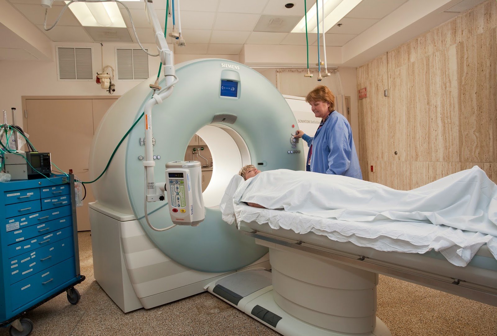
I must mean UFO or Unidentified Flying Object? You remember the movie Close Encounters of the Thrid Kind? Spielberg’s massive hit in 1977 following his release of the original Jaws. Back in those days UFO sightings were often in the news (or tabloids anyway) and this movie hit the sweet spot. It even helped launch the toy “Simon” which as it turns out was very similar to the multicolored, note-playing alien saucers featured in the movie – coincidence?


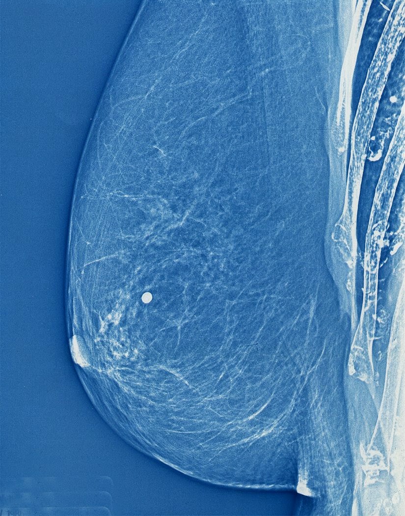
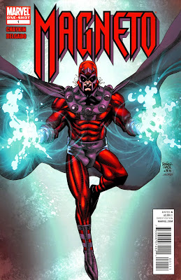
So, what the heck is UBO? Well, as it turns out the human body exhibits a variety of anatomical details in the ever so important Magnetic Resonance Imaging (MRI) scan that we have all learned to love (see our series on MRI and Carotid Stenosis). The majority of patients have similar anatomical features on imaging but some fall outside these normative patterns. When radiologists come across findings that are difficult  to interpret they will often refer to them as “Unidentified Bright Objects”. The challenge, of course, is that the radiologist needs to decide whether to label the anatomy in the image an “UBO” – essentially an image artifact – or “disease”.


This is where the rubber meets the road. Interpretation of MRI scans is work done by people, and, as with all jobs, the quality of performance varies. Therefore, the accuracy of the MRI exam is heavily dependent on the quality of the radiologists who interpret them. It is for this reason that the training a radiologist receives is crucial to his/her success. In addition, there is an important relationship that exists between the radiologist and the primary care physician as they have to balance indications of abnormality in MRI scans with the information provided by other techniques such as the clinical exam. A successful diagnosis relies on a good team effort. 


Go Team!


Now for the fun part (see the rules here), using UBO in a sentence by the end of the day:

Serious: Went for my MRI today. Told me that the UBO on imaging was just an artifact. Nothing to worry about. Phew!

Less serious: Hey Bob, did you hear on the news the report of another UBO hovering over farmer John’s field last night?  Or was that UFO? I always get those two mixed up…


Listen to UB [4] 0’s Red Red Wine to decompress and…


…I’ll see you in the blogosphere.


Pascal Tyrrell

MiWord of the Day Is… Magnet!

Who hasn’t thought of having Magneto’s powers? No? Maybe you should watch this Magneto trailer for a refresher. 


Ok, now that we all want to be Magneto (secretly at least) what is it that is so appealing with having the power of magnetism? Bill Nye the Science Guy explains it very well in this clip. Have a gander.


In a nutshell, magnetism is a physical phenomena that consists of a field of energy created by “magnets” that attracts or repels other objects. Magnets come in two major flavors: permanent magnets made of materials (such as iron) and electromagnets – the strongest and most widely used in medical imaging. 


Interestingly, it is the sum of the magnetic fields of individual electrons that is responsible for all the fun (see quantum mechanics). In the case of electromagnetism the electric current in a wire produces a magnetic field in the same direction of the current. In the case of a permanent magnet it is the magnetic fields of the naturally occurring electrically charged particles of the atoms that make up the material (iron for example) that are responsible. However, for there to exist a force strong enough to attract or repel another object all of its magnetic ions must have their magnetic fields aligned and contributing to the net magnetization. This is how you can magnetize a needle when stroking it in a uniform directional way with a permanent magnet.


Magnetism is to MRI what radiation is to X-rays. The strength of magnets is measured in gauss and Tesla units. There are 10,000 gauss to a Tesla and the earth’s magnetic field is one half of a gauss. Today most clinical MRIs use superconducting magnets whose strength range up to 4 Tesla! Experimental MRIs can run up to 10 Tesla. Now that is more Magneto’s speed.


The powerful magnets allow for better spacial resolution allowing for better sensitivity of the image. However, all this magnetic strength comes at a cost: the production of chemical shift artifacts – ghosts of things that are not really there. This is why we have radiologists to make sense of it all.


OK. Now you are asking what the heck. Magneto in the X-Men movie was able to rip out the iron from a human so why doesn’t an MRI? Great question. Iron found in the human body is mostly found as ferritin (a type of iron oxide) and is NOT magnetic. The iron in hemoglobin is also NOT magnetic. Bummer. So how does Magneto do it? Well either the movie is not scientifically correct (now that would be a shocker) or possibly he could be drawing on magnetite (another iron oxide) that is magnetic and has been found in trace amounts in the blood and brain. It is so little though that it does not cause any concern for MRI. Oh well, so much for Magneto…

Now for the fun part (see the rules here), using magnet in a sentence by the end of the day:

Serious: Hey Bob, did you know that early MRI machines used permanent magnets?

Less serious: Went for my MRI today. Told them I was worried the MRI would rip all the iron out of my blood like in X-Men. They didn’t even know who Magneto was. Whaaaat?!!

OK, listen to Magnetic by Traphik to decompress and I’ll see you in the blogosphere…

Pascal Tyrrell


MiWord of the Day Is… Xeroradiography!

Who hasn’t done some creative photocopying at some point in their lives? I certainly do NOT condone this type of activity (very naughty) but would you believe me if I were to tell you that for a long while mammography made use of photocopy technology? Yes, I realize this sounds a little funny. Let me explain.


In the 1970s medicine made the association between heavy exposure to radiation for TB and thyroid treatments and the appearance of breast cancer three decades later. A reevaluation of the effects of radiation ensued and a call for ways to minimize exposure to ionizing radiation was made to the industry.


One of the first to answer that call was the radiologist John Wolfe from Detroit Receiving Hospital who in 1966 reported on the advantages of coupling photocopy technology with mammography. Xerox corporation jumped on the idea and developed a commercial unit in 1971 and “xeroradiography” was born! Basically, film from traditional x-ray imaging (yes back then they still used film!) was replaced with a selenium coated aluminum plate that was prepared for the exposure by being electrically charged. The result was that only a short burst of radiation (shorter exposure time means lower dose of radiation) was required to produce a very high quality image.


These xerox mammograms dominated the industry for over 20 years until new technology was developed more recently that provided even finer images with even less radiation. Cool.


Now for the fun part (see the rules here), using Xeroradiography in a sentence by the end of the day:


Serious: Hey Bob, did you know that mammograms produced using xeroradiography were blue? 


Less serious: My friend Jane was scheduled for a mammography. Having heard of xeroradiography reading the MiVIP blog she decided to DIY at her office. Problem was the print kept coming out black and white instead of blue from the Xerox machine…


OK, watch the Copy Cat trailer to decompress (or not?!!!) and I’ll see you in the blogosphere…


Pascal Tyrrell

MiWord of the Day Is… Mesopotamia!

You are thinking about pursuing studies in medicine. You have enrolled in all the necessary courses at school to qualify you for the grueling application process and you are actively looking for volunteer opportunities. So why the need to be active in your community?


Today, I want to talk a little about the history of medicine. Around 3000 BC (and no I was not alive then if you are wondering) the middle east was a hotbed for civilizations who were in transition from being mainly nomadic to more settled. This “land between the rivers” – Mesopotamia – was ruled by many successive great kingdoms including the Akkadian, Babylonian, and Assyrian empires. Thanks to many archaeological and written remains we have discovered that healing practices indeed existed and were established during these times.


Mesopotamian medicine was predominantly religious and was delivered by a team of healers: the seers who would diagnose based on divination, the exorcists who would expel demons, and finally the physician priests who actually treated the sick mostly with charms, drugs, and some surgical procedures.  OK, so this intensely codified approach (which meant very little opportunity for discussion)  to healing that dominated the Mesopotamian kingdoms would not be able to adapt or improve much over time and would ultimately not contribute much to the Greek rational medicine that would come a later and evolve into today’s medicine. 


So why is it important? For two reasons:


Firstly, by understanding the history of medicine you will better appreciate the importance of your role as a physician in your community – regardless if you are a primary care physician on the front line or a radiologist who works in the back ground. What is important is to feel connected and part of your community.


Secondly, it is interesting to see that though Mesopotamian medicine recognized very early on that factors like cold, alcohol, and unhygienic conditions affected health, they were enable to advance and evolve their medicine as Ancient Greece did through ongoing experimentation and discussion. Moral of the story? Medical research rocks!


Do you remember the Babylon 5 series? It came many, many, many years later! Have a peek to decompress and…


… I’ll see you in the blogosphere.


Pascal Tyrrell


MiWord of the Day Is… CAT scan!

A what scan? I am actually a cat guy myself. Not to say I don’t love dogs but if I had to make a choice…


I just finished reading a fantastic book by David Dosa entitled “Making Rounds With Oscar”. The premise of the book is a story about an extraordinary cat but the subject matter is very serious –  dementia and end-of-life care in the elderly. Have a gander.


So what the heck is a cat scan and what does it have to do with medical imaging? 


CT scans – also referred to as computerized axial tomography (CAT) – are special X-ray tests that produce cross-sectional images of the body using X-rays and a computer. CT was developed independently by a British engineer named Sir Godfrey Hounsfield and Dr. Alan Cormack and were jointly awarded the Nobel Prize in 1979. Yes, more Nobel prize winners…


In a nutshell, x-ray computed tomography:


– uses data from several X-ray images of structures inside the body and converts them into 3D pictures – especially useful for soft tissues.


– emits a series of narrow beams through the human body, producing more detail than standard single beam X-rays.


– is able to distinguish tissues inside a solid organ. A CT scan is able to illustrate organ tear and organ injury quickly and so is often used for accident victims.


– is analyzed by radiologists.


Unfortunately, unlike MRI scans, a CT scan uses X-rays and therefore are a source of ionizing radiation.


Now for the fun part (see the rules here), using CAT Scan in a sentence by the end of the day:

Serious: Hey Bob, did you know that the recorded image of a CAT Scan is called a tomogram? 

Less serious: My GP suggested that howling at the moon at night is not normal behavior and he wants to send me for a CAT scan. What? No way, I’m allergic to cats…

OK, listen to Cat Stevens to decompress and I’ll see you in the blogosphere…


Pascal Tyrrell