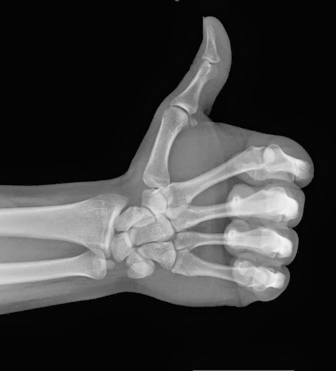
Yup! Want some of that. Not only is Superman cool but he has x-ray vision. Unbelievable. Or is it? Radiologists have the same x-ray vision but without the Spandex suit – or at least they don’t wear it to work that I am aware of.
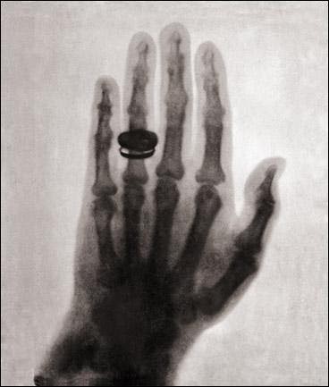
The word of the day is x-ray. You have already successfully used “Roentgen” in casual conversation last week (don’t know what I am talking about? See Mi Word of the Day Is… Roentgen!) and today I will talk a little about what Roentgen was first in measuring and describing – x-rays.
Let’s say you are in your lab and you are working with passing electrical discharges through vacuum tubes – a typical Saturday afternoon activity with friends. As chance would have it your little sister’s barium salts paintings happen to be drying near-by and you notice a faint glow emanating from them every time you run your experiments. No matter how much you try to block any light coming from your vacuum tubes the glow persists. What? That’s odd. How’s that happening? Well my friend, you have just crossed over into the Twilight Zone (awesome old tv series) and discovered a form of electromagnetic radiation.
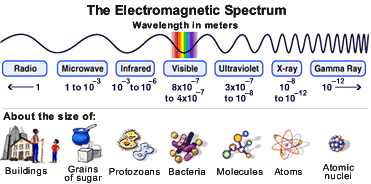 Visible light is but a very small part of the electromagnetic spectrum. Moving from visible light to longer wavelengths and lower frequencies we find infrared (keeps food warm at restaurants), microwaves (to warm your pizza pop) and radio (not the one streamed through the internet!).
Now if you move in the opposite direction from visible light you find shorter wavelengths with higher frequencies starting with ultraviolet (what helps you get that summer tan), x-rays (word of the day), and finally gamma rays (topic for another day!). So x-rays are about the size of atoms and radio waves the size of buildings. Crazy. I think what is surprising is that with the naked eye we “see” so little and yet so much (philosophy anyone?).
So, x-rays are short wavelength, high frequency, high energy electromagnetic radiation that is able to penetrate some substances more easily than others. For example, they penetrate flesh more easily than bone, and bone more easily than lead. Thus they make it possible to see bones within flesh and a bullet embedded in bone. The ability of X rays to penetrate depends on their wavelength and on the density and thickness of the substance being scanned.
2- You have to use the word in a sentence by the end of the day. No need to use it in the correct context – actually out of context is more fun and elicits a more entertaining response!
Serious: Hey Frank, did you know the radiation you received during your chest x-ray last week was actually “soft” x-rays? Ones with shorter wavelengths and more penetrating power are used for scanning archaeological artifacts.
 Less serious: Frank! Dude, I got them! My x-ray specs just came in the mail. Let’s go the beach…
See you in the blogosphere,
Pascal Tyrrell