The Adventures of Alana the Newbie Researcher

Timeline of my ROP experience

Stroke. Atherosclerosis. Carotid stenosis. MRI. Endarterectomy. To some people, these words are, well, just words. But for others, these few phrases could change the course of their lives. During my time in the Research Opportunity Program at the Department of Medical Imaging, I was able to learn all about carotid disease and the current status of carotid imaging modalities. As an added bonus, I did quite a bit of research on the impact that stenosis has on patient care and how new and exciting advances in medical imaging could affect it.


Over the course of four months, I went from being a huge research newbie to becoming an experienced research … trainee. I was introduced to the world of journal article writing, starting from doing elementary literary research all the way to drafting and writing a review article, which was completed entirely from the comfort of my beloved computer. With the help of Doctor Eli Lechtman and my ROP partner Helena Lan (if you haven’t seen her post on her experience you should check it out), we were able to put together 2 review articles that complement one another and will hopefully pose as a good foundation for future research.


Now, you must be thinking, “wow, this sounds like an extremely exciting summer; how could it possibly be better?” Well, my dear readers, it gets even better. One of the greatest things about being a part of this department is the community. The people here are all friendly, wonderful, and welcoming. From having an outing at Centre Island to letting us operate an uItrasound machine (thanks Modele!), I really felt like I was a part of “the gang.” What’s more, it was nice to have someone remember my name when I arrived in the morning, especially when I’m used to being my nameless self in the vast sea of students of our first year lectures (Convocation Hall, anyone?).


When I first sat down to write this post, I couldn’t decide what would be the best way to describe my time doing my ROP. And that’s when it hit me: it was an adventure! I may not have explored unknown lands or survived any near-death experiences, but there was not one day that went by where I wasn’t learning something new. Each day was a new experience, an opportunity to understand something I probably haven’t even heard of before. It still surprises me right now, as I’m writing this post, how much I have learned throughout this program, and the fact that I was able to experience something like this. I would have never thought I could write a review article and do the things I’ve done in only my first year of university. All in all, this was a great experience, and I encourage everyone to try out something like this!


Thanks for reading this, y’all! And if you just scrolled to the bottom of this post and thought “tl;dr,” you can still check out my infographic timeline (there’s pretty pictures)! Also I’d like to give a shoutout to my awesome Professor, Dr. Pascal Tyrrell (check out his latest post), as well as all the lovely members of the Medical Imaging department at U of T for making my ROP experience the way it turned out to be.


Remember to make every day an adventure,


Alana Man 🙂

Helena Lan: Summer 2014 ROP

Helena Lan Summer 2014 ROP

What is research like? If you had asked me this
question several months ago, I would have answered, “You wear a lab coat and
goggles while mixing chemicals or observing organisms. Hopefully something
interesting will happen, so that you get to publish your findings!” Well, after
participating in the Research Opportunity Program (ROP) at the University of
Toronto, I discovered that medical imaging research is more than just
pipetting, and is all the more exciting!
 
So what kind of research is conducted in the medical imaging world? For my ROP, the objective of my project was to evaluate the roles of the non-invasive imaging modalities for diagnosing carotid stenosis. Hence, I engaged in online literature research of the various imaging techniques for assessing this disease. In this process, I also learned to use Zotero to manage all my references, which provides an easy way to generate a bibliography (when the software doesn’t crash every time you open it). After gathering all the pertinent information, I then put together a review article suggesting how a change in the current imaging approach could potentially improve clinical outcome. Who knew a report could be compiled without doing the lab grunt work?     
 
Wait, so this is all a radiologist does? Sitting in front of a computer and typing all day? Of course not! During our time at Sunnybrook Hospital, we got the chance to chat with a radiologist and discovered that she could decide whether patients should be released after taking a look at their diagnostic images. Pretty powerful, eh? That’s not all. We also found out how radiologists identified any abnormalities in patients, as we had the opportunity to work with the VesselMass software which allowed for the delineation of the lumen and vessel wall of arteries on MRI images. Oh, and did I mention we observed an MRI and an ultrasound examination of the carotid arteries, and even got to perform an ultrasound scan ourselves. Super cool!
 
Still craving for more of my ROP experience? Check out my timeline infographic! You will find all the things I learned and all the fun I had there. Last but not least, I’d like to shout out a big THANK YOU to Prof. Pascal Tyrrell and Dr. Eli Lechtman, who guided us every step of the
way. Also, I’m very grateful to Dr. Alan Moody for including us in his research program at Sunnybrook, as well as other members of the VBIRG group who gave us the chance to
participate in various activities. My summer would not have been this fun and meaningful without all of your help!
 
Have fun researching,


Helena Lan
 

A YSP Student Perspective: MRI and Carotid Artery Disease

Hershel Stark, MED YSP 2014 Student

Throughout the month of July, I participated in a research program with the Division of Teaching Laboratories within the Faculty of Medicine at the University of Toronto. I was assigned to work with Prof. Pascal
Tyrrell and the Department of Medical Imaging, and spent the majority of my time with the Vascular Biology Imaging Research Group (VBIRG) at Sunnybrook Research Institute. I would like to discuss my experiences, what I gained from the program, and how I can take those skills with me into the future.


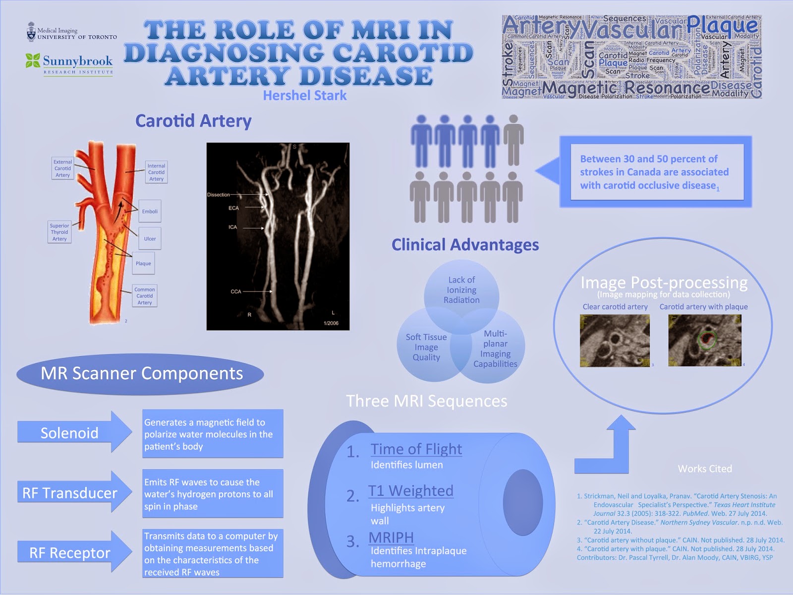
Essentially, the program was composed of presentations and shadowing opportunities in which I was introduced to various imaging modalities used in both the clinical and research fields. I primarily studied MR imaging, but was nevertheless exposed to other modalities including ultrasound and CT.  Towards the end of the program, I had two principal objectives: to present my experiences to the VBIRG group and to design an infographic for displaying. Below is a copy of my infographic:

Notwithstanding the abundance of knowledge I gained from studying the subject content, I acquired a variety of essential research skills by partaking in the program. Shadowing proficient researchers as they collected
and analyzed data provided me with a thorough insight of a researcher’s methods and techniques. The researchers that I worked with appropriately explained their individual roles on the research team, which led to my understanding of the significance of collaboration in scientific and medical research.
One last aspect of the program that I would like to address is the daily workshops that were conducted by two instructors from the Division of Teaching Laboratories, Jastaran Singh and Jabir Mohamed. Each of these brief workshops focused on an important general topic relevant to research in general, ranging from discussing common scientific practices to elaborating on literary research. I believe that the combination of skills and knowledge that I obtained from all elements of the program will be useful in my potential
research career in university.
 Lastly, I would like to take this opportunity to formally thank all of those that contributed to making the program a truly enjoyable and intellectually stimulating experience. I would like to extend my gratitude to
Dr. Alan Moody and the members of the VBIRG group for allowing me to shadow their research projects, as well as to Prof. Pascal Tyrrell and the Department of Medical Imaging at U of T for constructing the program and offering much assistance in the formation of my infographic. Finally, I’d like to thank Dr. Chris Perumalla and the Division of Teaching Laboratories in the Faculty of Medicine at U of T for formulating the research module of the Youth Summer Program, and Jastaran Singh and Jabir Mohamed for providing guidance as instructors throughout the program.
Best of luck in all of your future endeavours,
Hershel Stark