Who hasn’t done some creative photocopying at some point in their lives? I certainly do NOT condone this type of activity (very naughty) but would you believe me if I were to tell you that for a long while mammography made use of photocopy technology? Yes, I realize this sounds a little funny. Let me explain.
In the 1970s medicine made the association between heavy exposure to radiation for TB and thyroid treatments and the appearance of breast cancer three decades later. A reevaluation of the effects of radiation ensued and a call for ways to minimize exposure to ionizing radiation was made to the industry.
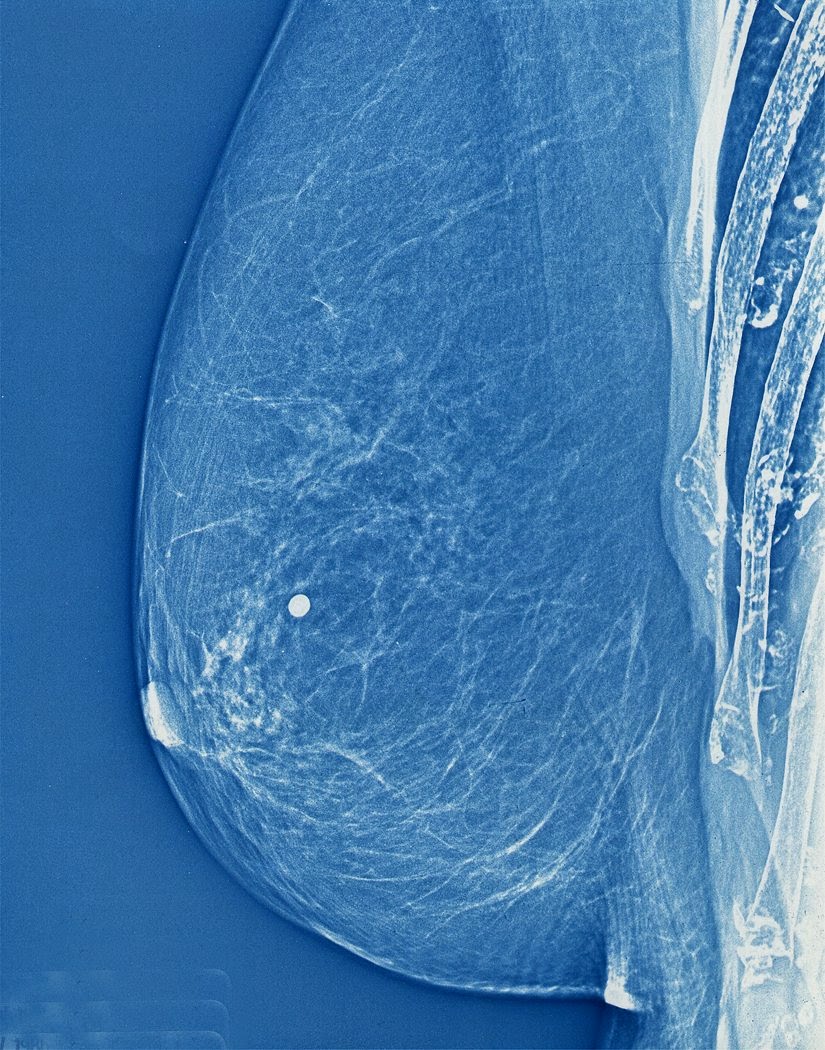 One of the first to answer that call was the radiologist John Wolfe from Detroit Receiving Hospital who in 1966 reported on the advantages of coupling photocopy technology with mammography. Xerox corporation jumped on the idea and developed a commercial unit in 1971 and “xeroradiography” was born! Basically, film from traditional x-ray imaging (yes back then they still used film!) was replaced with a selenium coated aluminum plate that was prepared for the exposure by being electrically charged. The result was that only a short burst of radiation (shorter exposure time means lower dose of radiation) was required to produce a very high quality image.
These xerox mammograms dominated the industry for over 20 years until new technology was developed more recently that provided even finer images with even less radiation. Cool.
Now for the fun part (see the rules here), using Xeroradiography in a sentence by the end of the day:
Serious: Hey Bob, did you know that mammograms produced using xeroradiography were blue?
Less serious: My friend Jane was scheduled for a mammography. Having heard of xeroradiography reading the MiVIP blog she decided to DIY at her office. Problem was the print kept coming out black and white instead of blue from the Xerox machine…
OK, watch the Copy Cat trailer to decompress (or not?!!!) and I’ll see you in the blogosphere…
Pascal Tyrrell
MiWord of the Day Is… Xeroradiography!