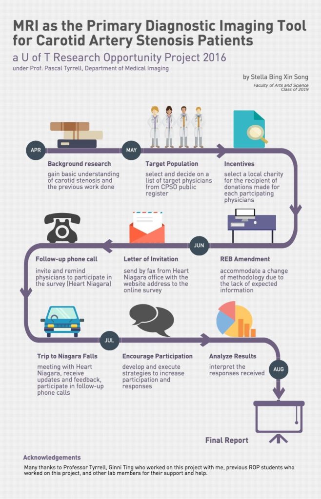
MiWord of the Day Is… Magnet!

Who hasn’t thought of having Magneto’s powers? No? Maybe you should watch this Magneto trailer for a refresher. 


Ok, now that we all want to be Magneto (secretly at least) what is it that is so appealing with having the power of magnetism? Bill Nye the Science Guy explains it very well in this clip. Have a gander.


In a nutshell, magnetism is a physical phenomena that consists of a field of energy created by “magnets” that attracts or repels other objects. Magnets come in two major flavors: permanent magnets made of materials (such as iron) and electromagnets – the strongest and most widely used in medical imaging. 


Interestingly, it is the sum of the magnetic fields of individual electrons that is responsible for all the fun (see quantum mechanics). In the case of electromagnetism the electric current in a wire produces a magnetic field in the same direction of the current. In the case of a permanent magnet it is the magnetic fields of the naturally occurring electrically charged particles of the atoms that make up the material (iron for example) that are responsible. However, for there to exist a force strong enough to attract or repel another object all of its magnetic ions must have their magnetic fields aligned and contributing to the net magnetization. This is how you can magnetize a needle when stroking it in a uniform directional way with a permanent magnet.


Magnetism is to MRI what radiation is to X-rays. The strength of magnets is measured in gauss and Tesla units. There are 10,000 gauss to a Tesla and the earth’s magnetic field is one half of a gauss. Today most clinical MRIs use superconducting magnets whose strength range up to 4 Tesla! Experimental MRIs can run up to 10 Tesla. Now that is more Magneto’s speed.


The powerful magnets allow for better spacial resolution allowing for better sensitivity of the image. However, all this magnetic strength comes at a cost: the production of chemical shift artifacts – ghosts of things that are not really there. This is why we have radiologists to make sense of it all.


OK. Now you are asking what the heck. Magneto in the X-Men movie was able to rip out the iron from a human so why doesn’t an MRI? Great question. Iron found in the human body is mostly found as ferritin (a type of iron oxide) and is NOT magnetic. The iron in hemoglobin is also NOT magnetic. Bummer. So how does Magneto do it? Well either the movie is not scientifically correct (now that would be a shocker) or possibly he could be drawing on magnetite (another iron oxide) that is magnetic and has been found in trace amounts in the blood and brain. It is so little though that it does not cause any concern for MRI. Oh well, so much for Magneto…

Now for the fun part (see the rules here), using magnet in a sentence by the end of the day:

Serious: Hey Bob, did you know that early MRI machines used permanent magnets?

Less serious: Went for my MRI today. Told them I was worried the MRI would rip all the iron out of my blood like in X-Men. They didn’t even know who Magneto was. Whaaaat?!!

OK, listen to Magnetic by Traphik to decompress and I’ll see you in the blogosphere…

Pascal Tyrrell


MiCUP… Runneth Over?

An interesting quotation from the Hebrew bible. Basically it means that I have sufficient for my needs and I am good with that. So, where am I going with this you ask? Well, let me introduce you to my program MiCUP – Medical imaging Collaborative Undergraduate Program. 


The goal of the program is to bring together students from the faculty of Arts and Sciences and my faculty (Medicine) to learn about medical research in the world of medical imaging. I have a sprinkling of students every term from various programs such as Research Opportunity Program, Independent Studies, Youth Study Program, and MiVIP. It is only a modest number of students BUT provides ample brain power to get some really cool research done. My cup certainly runneth over. 


Have a look below at the timelines from my two recent ROP students.


Great work Kevin and Sylvia!!!


See you in the blogosphere,


Pascal Tyrrell


Kevin Chen ROP F/W 2014

Sylvia Urbanik F/W 2014

Basic Functions and Why You Should Know About Them

No, I did not say “bodily functions”. That is discussed in another blog. We’re talking math today. 


So, my son was doing his homework the other night and yelled out from his room:”Daaaadddyyyy!!! Do you know what a parabola is?” For those of you who do not have teenage children this is code for “can you help me with my homework”. After reliving a few high school memories that came along with the word “parabola” I wondered over to his room to see what the latest homework challenge was going to be…


When helping my kids with their homework, I often think of how important and still relevant some of the basic math is we learnt in high school. I would like to talk a little about basic functions and how they are still used well after you have handed in your last math homework assignment.


Many (most?) scientific laws are expressed as relations between two or more variables – often physical quantities. Next comes the chicken or the egg conundrum. Were the results from an experiment used to formulate “empirical laws” or did we use existing knowledge and math to come up with new theories – that we will invariably later have to test. Welcome to the world of research!


If two variables are related in such a way that one of them (the dependent or response variable) is determined when the other is known (the independent or explanatory variable), then there exists what is termed a functional relationship between the variables.


y = f(x)


For example the relationship of height to weight in humans. In general, the taller we are the heavier we get. This results in what is called a straight-line relationship.


But not all relationships are linear. How about if we were to throw a ball up into the air and measure it’s trajectory? It would look a little like the picture on the left.


Although initially the value of the height of the ball increases with time, there comes a point when the ball stops rising and starts to fall back down to earth. The resulting curve is called – you guessed it – a parabola.


The math functions for the parabola and that of the straight line are actually related. Yes, I am serious! They both belong to the family of math functions called polynomials. In my next posts I will talk a little about how we describe these functions and how we can put them to work for us in the world of medical research.


For now, decompress watching this hilarious movie trailer Biloxi Blues which is all about basic training (you can now relate) and…


… I’ll see you in the blogosphere,


Pascal Tyrrell

MiWord of the Day Is… Xeroradiography!

Who hasn’t done some creative photocopying at some point in their lives? I certainly do NOT condone this type of activity (very naughty) but would you believe me if I were to tell you that for a long while mammography made use of photocopy technology? Yes, I realize this sounds a little funny. Let me explain.


In the 1970s medicine made the association between heavy exposure to radiation for TB and thyroid treatments and the appearance of breast cancer three decades later. A reevaluation of the effects of radiation ensued and a call for ways to minimize exposure to ionizing radiation was made to the industry.


One of the first to answer that call was the radiologist John Wolfe from Detroit Receiving Hospital who in 1966 reported on the advantages of coupling photocopy technology with mammography. Xerox corporation jumped on the idea and developed a commercial unit in 1971 and “xeroradiography” was born! Basically, film from traditional x-ray imaging (yes back then they still used film!) was replaced with a selenium coated aluminum plate that was prepared for the exposure by being electrically charged. The result was that only a short burst of radiation (shorter exposure time means lower dose of radiation) was required to produce a very high quality image.


These xerox mammograms dominated the industry for over 20 years until new technology was developed more recently that provided even finer images with even less radiation. Cool.


Now for the fun part (see the rules here), using Xeroradiography in a sentence by the end of the day:


Serious: Hey Bob, did you know that mammograms produced using xeroradiography were blue? 


Less serious: My friend Jane was scheduled for a mammography. Having heard of xeroradiography reading the MiVIP blog she decided to DIY at her office. Problem was the print kept coming out black and white instead of blue from the Xerox machine…


OK, watch the Copy Cat trailer to decompress (or not?!!!) and I’ll see you in the blogosphere…


Pascal Tyrrell

MRA Is More Cost-Effective than the Current Strategy for Testing > 70% Carotid Stenosis

ROP Research Forum March 5th, 2015 – Kevin Chen


Kevin Chen is a 2nd year research opportunity program student studying neuroscience and
physiology in the University of Toronto Life Sciences Program. His main goal is to get into
medical school and to enjoy the UofT experience as much as possible!

Kevin’s ROP project consisted
of cost effectiveness analysis modeling to explore whether MRA could be a
cost-effective measure in testing and treating patients with carotid stenosis.
Preliminary results showed that in a subset of the population at risk for
carotid stenosis (> 70 %), MRA was dominant over the current strategy of testing with
Doppler Ultrasound. By reducing MRA scan time and by tailoring MRA sequences we believe it possible to extend these findings to include a larger sub-population (> 50 %). More to follow…

Well done, Kevin!

See you in the blogosphere,

Pascal Tyrrell

Research Opportunity Program: Cost Effectiveness and Imaging Carotid Stenosis

ROP Research Forum March 5th, 2015 – Sylvia Urbanik

Sylvia is a second year student studying cell and molecular biology. She is currently finishing up her Research Opportunity Program that spanned the fall and winter semesters, and also recently represented us at the March 5th ROP research forum along with her partner and predecessors.

 
Her project dealt with cost-effectiveness analysis techniques for diagnostic imaging modalities, with a specific focus on carotid artery stenosis. She explored the different types of analyses used in assessing cost effectiveness. She examined how factors such as sensitivity and specificity of diagnostic tests (imaging modalities in this case) can affect the analysis, and conducted literature searches in order to find these variables in order to incorporate them into a cost effectiveness model.​

Well done, Sylvia!

Stay tuned, next Kevin Chen will be modeling with TreeAge…

See you in the blogosphere,

Pascal Tyrrell

Research Opportunity Program: Imaging Carotid Stenosis

ROP Research Forum March 5th, 2015 – Helena Lan

Helena is a second-year student pursuing a specialist in Pharmacology and Toxicology at the University of Toronto. Participating in ROP299 this past summer (see here) has opened many doors for her. She is currently assisting Pascal on a systematic review on research methodology and biostatistics in medical imaging, working as a research assistant for a medical education study, participating in the research abroad program at Karolinska Institutet this summer… and recently represented us at the March 5th, 2015 ROP research forum!

Helena examined the technical aspects and information provided by two major imaging modalities, MRI and ultrasound, for diagnosing carotid stenosis. She suggests that MRI holds great promise to serve as a cost effective test for carotid stenosis as well as a tool for assessing vessel health and plaque composition that would provide important information for patient management decisions.


Stay tuned, next Sylvia Urbanik will be talking about cost effectiveness…

See you in the blogosphere,

Pascal Tyrrell

Research Opportunity Program: What Is Carotid Stenosis and How Are We Treating Affected Patients?

ROP Research Forum March 5th, 2015 – Alana Man

Alana Man is a second year University of Toronto student pursuing a specialist in Bioinformatics and Computational Biology and a major in Immunology. She was a Research Opportunity student last summer with MiVIP (see here) and recently represented us at the March 5th, 2015 ROP research forum.

Alana’s project focused on care for patients with carotid stenosis in Toronto, Ontario, Canada. She looked at the different factors contributing to access to diagnostic testing such as procedure costs, wait times, and the differences between available imaging modalities and explores whether MRI could be the diagnostic test of choice over DUS for people at increased risk of carotid stenosis.

Well done, Alana!


Stay tuned, next Helena Lan will be comparing MRI and DUS…


See you in the blogosphere,


Pascal Tyrrell

MiWord of the Day Is… PET scan!


Peanuts. What a great story. The most popular and influential comic strip in history. Snoopy was my first stuffed animal growing up. He still lives with my parents. So what is a PET scan anyway? I don’t recall ever seeing the picture above in any of the Peanuts cartoon strips.


Positron emission tomography (PET) is somewhat of a special medical imaging modality in that it brings together two different technologies from different times. Let me explain. Back in the early 1930s, George Hevesy was a young Hungarian physicist who developed biologically safe and useful radioactive tracers that could be ingested or incorporated into the body in some way.  Physicians would then manually locate where these radioactive tracers had gone in the body by using a Geiger counter at first and then later using special cameras (Kuhl‘s photoscan) to produce a crude emission image. 


So, how do we get cool pictures like these ones?  Well we would have to wait another 25 years after the development of radioactive tracers by Hevesy for the start of construction of instruments able to not only detect these radioactive sources in the body but to produce tomographic pictures.


It won’t be until the mid 1970s that PET – as we know it today – would be born. Essentially, a patient receives a emissions scan (PET) and a CT (we talked about that here) or MRI (we talked about that here) scan at the same time. The two scans are then merged together thanks to highly specialized computers (see the pictures in the middle frames). Voila! PET. 


PET is both a medical and research tool. Most often used in clinical oncology (medical imaging of tumors and the search for metastases), it is also important in clinical diagnosis of certain diffuse brain diseases such Alzheimer’s disease and other types of dementia.


Relax your brain a little listening to Radioactive by Imagine Dragons and don’t forget the fun part (see the rules here), using PET scan in a sentence by the end of the day:

Serious: Hey Bob, did you know that much of the success of the PET scan is due to the development of the radiopharmaceutical FDG (deoxyglucose) that lead the way to the characterization of Parkinson’s and Huntington’s disease?

Less serious: I can’t believe they developed yet another PET scan. Wasn’t the CAT scan enough?

See you in the blogosphere,

Pascal Tyrrell


Walk Like an Egyptian!

So, in my last post I talked a little about Mesopotamian medicine (see here). I am certain many of you were thinking: “What? Should he not be talking about ancient Egypt?”. Well, of course, you are right – kind of… 


Egypt rose under the pharaohs during the same period as the Mesopotamian kingdoms (from about 3000 BC). They were known for their crazy ambition and technological prowess. Their medicine was very similar to that of the Mesopotamians in that it was influenced strongly by superstition  and religious beliefs. They too had three types of healers: the swnu who practiced medicine, and, of course, the priests and the sorcerers…


One of the reasons that ancient Egyptian medicine had a greater influence on modern medicine was that they were very good at documenting and archiving their work. The Ebers papyrus (c. 1550 BC) was their principal medical document that measured over 20 meters long (it is a scroll after all) and is the oldest surviving medical book. 


The Egyptians believed we were all born healthy but were susceptible to disorders caused by demons or by intestinal putrefaction. So the importance of eating your fruits and veggies was started way long ago! They also compared our vascular network to that of the River Nile and its canals and, therefore, it was important to keep the flow free from obstructions (see here for another interesting comparison!). Though they did not appreciate vascular plaques (atheroma) at the time they had already started to  figure out the importance of a healthy vasculature. Cool! 


As with Mesopotamia, Egypt’s powerful governance created a good environment for organized medical practice. However, because both regimes were highly codified (implying many strict rules based on religion and superstition that did not allow for discussion and experimentation) it will not be until ancient Greece that the roots of modern medicine will take hold.


Dance around your living room (in private if you must) to Walk Like an Egyptian by The Bangles in order to decompress and…


… I’ll see you in the blogosphere.


Pascal Tyrrell