Some R&R with UofT YSP and Elizabeth Lehner…

Elizabeth Lehner – YSP 2015


Maybe not all rest and relaxation but certainly radiology and rheumatology! Here is a great example of why collaboration between disciplines is so important in medicine. Elizabeth recently graduated from Iroquois Ridge High School and will be a new University of Toronto student this fall. See her post below. 


Great job Elizabeth!!!

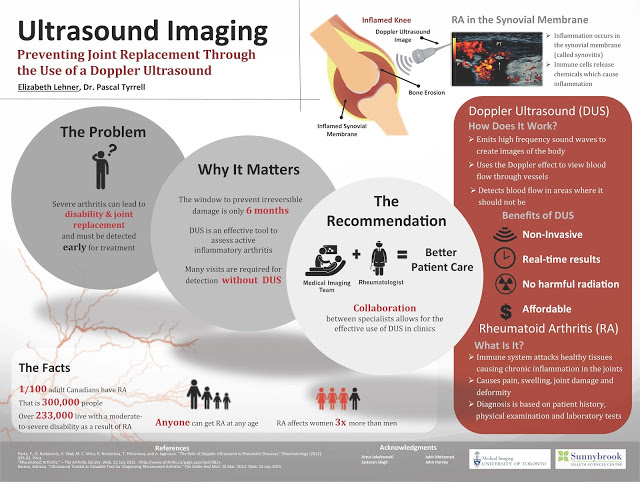
Many people are familiar with the word arthritis. This is probably because one in six Canadians aged 15 years and older report having arthritis. Rheumatoid Arthritis is a specific form of arthritis that unfortunately can lead to severe disability and joint replacement. 


Over the past several weeks, I participated in the 2015 YSP research program with the Division of Teaching Laboratories within the Faculty of Medicine at the University of Toronto and had the opportunity to look more closely at Rheumatoid Arthritis and ways to better diagnose this debilitating disease.


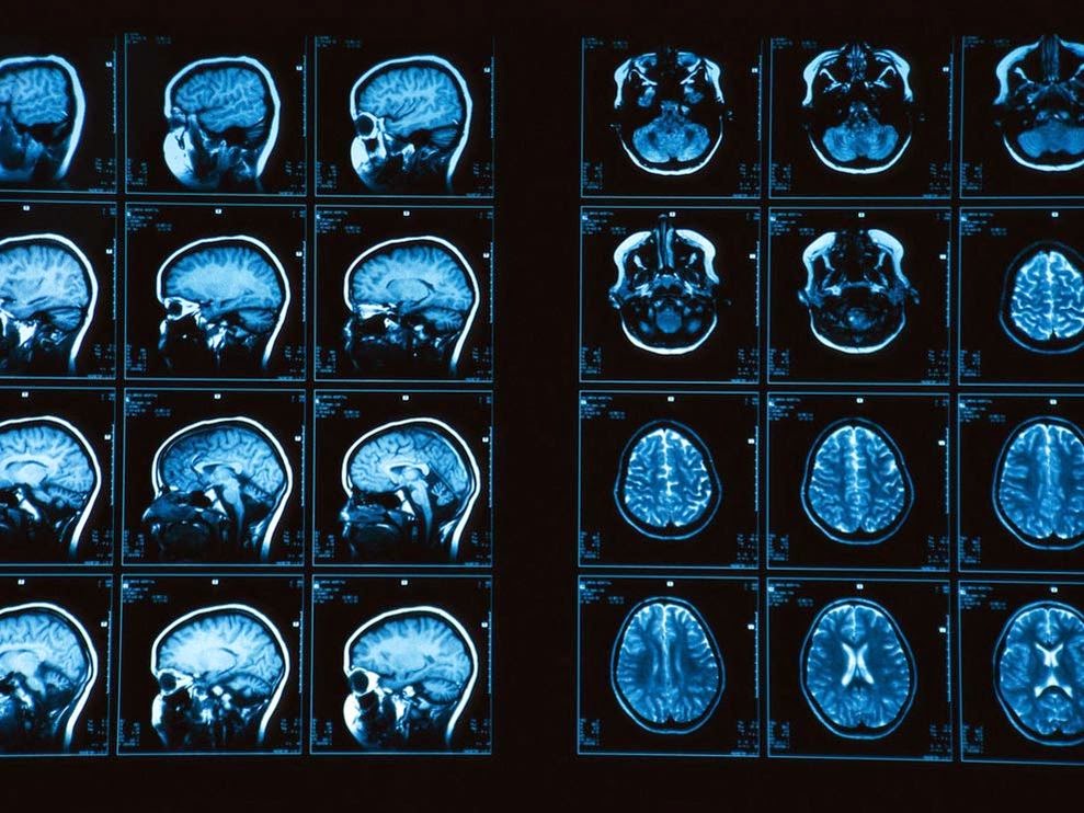
Under the supervision of Prof. Pascal Tyrrell and the Department of Medical Imaging at U of T, I was introduced to various imaging modalities including MRI machines, CT scanners and ultrasound machines. The work by Dr. Tyrrell was of particular interest given his studies on inflammation and the use of the various imaging modalities.


As part of this program I also participated in specific lab tasks including dissections and micropipetting and was exposed to clinical work such as suturing and operating an ultrasound machine. In addition, the program provided me with the opportunity to participate in daily workshops led by two instructors from the Division of Teaching Laboratories, Jastaran Singh and Jabir Mohamed. These workshops provided important overviews on a variety of topics relating to research that were very interesting.


The things I learned in this program provided me with a much better understanding of various research and medical issues that I think will be of use to me as I begin my studies at the University of Toronto this fall.
I would very much like to thank Prof. Pascal Tyrrell, Jastaran Singh and Jabir Mohamed for allowing me to be exposed to the various projects and for answering the many questions that I had during the program. Thank you!

 
Elizabeth Lehner

MiWord of the Day Is… Xeroradiography!

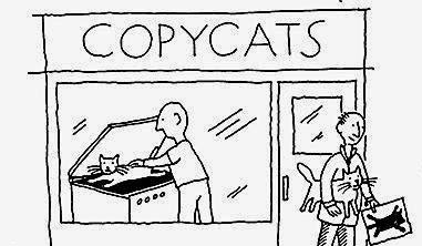
Who hasn’t done some creative photocopying at some point in their lives? I certainly do NOT condone this type of activity (very naughty) but would you believe me if I were to tell you that for a long while mammography made use of photocopy technology? Yes, I realize this sounds a little funny. Let me explain.


In the 1970s medicine made the association between heavy exposure to radiation for TB and thyroid treatments and the appearance of breast cancer three decades later. A reevaluation of the effects of radiation ensued and a call for ways to minimize exposure to ionizing radiation was made to the industry.


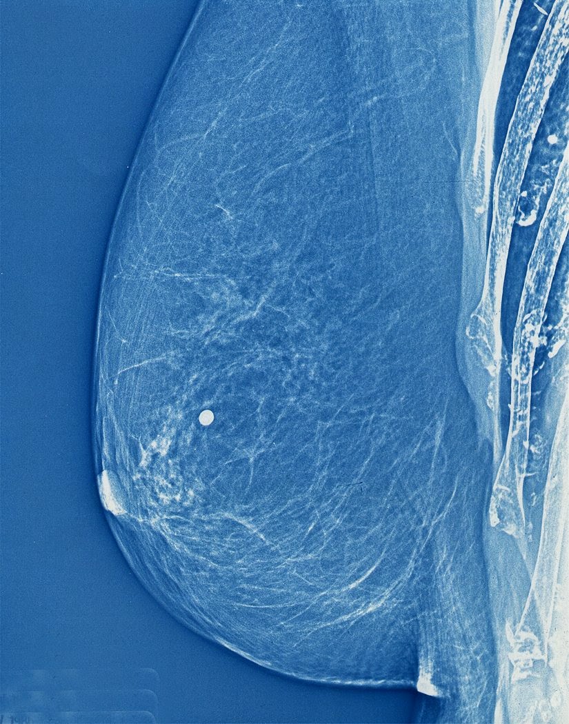
One of the first to answer that call was the radiologist John Wolfe from Detroit Receiving Hospital who in 1966 reported on the advantages of coupling photocopy technology with mammography. Xerox corporation jumped on the idea and developed a commercial unit in 1971 and “xeroradiography” was born! Basically, film from traditional x-ray imaging (yes back then they still used film!) was replaced with a selenium coated aluminum plate that was prepared for the exposure by being electrically charged. The result was that only a short burst of radiation (shorter exposure time means lower dose of radiation) was required to produce a very high quality image.


These xerox mammograms dominated the industry for over 20 years until new technology was developed more recently that provided even finer images with even less radiation. Cool.


Now for the fun part (see the rules here), using Xeroradiography in a sentence by the end of the day:


Serious: Hey Bob, did you know that mammograms produced using xeroradiography were blue? 


Less serious: My friend Jane was scheduled for a mammography. Having heard of xeroradiography reading the MiVIP blog she decided to DIY at her office. Problem was the print kept coming out black and white instead of blue from the Xerox machine…


OK, watch the Copy Cat trailer to decompress (or not?!!!) and I’ll see you in the blogosphere…


Pascal Tyrrell

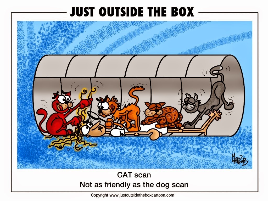
MiWord of the Day Is… CAT scan!

A what scan? I am actually a cat guy myself. Not to say I don’t love dogs but if I had to make a choice…


I just finished reading a fantastic book by David Dosa entitled “Making Rounds With Oscar”. The premise of the book is a story about an extraordinary cat but the subject matter is very serious –  dementia and end-of-life care in the elderly. Have a gander.


So what the heck is a cat scan and what does it have to do with medical imaging? 


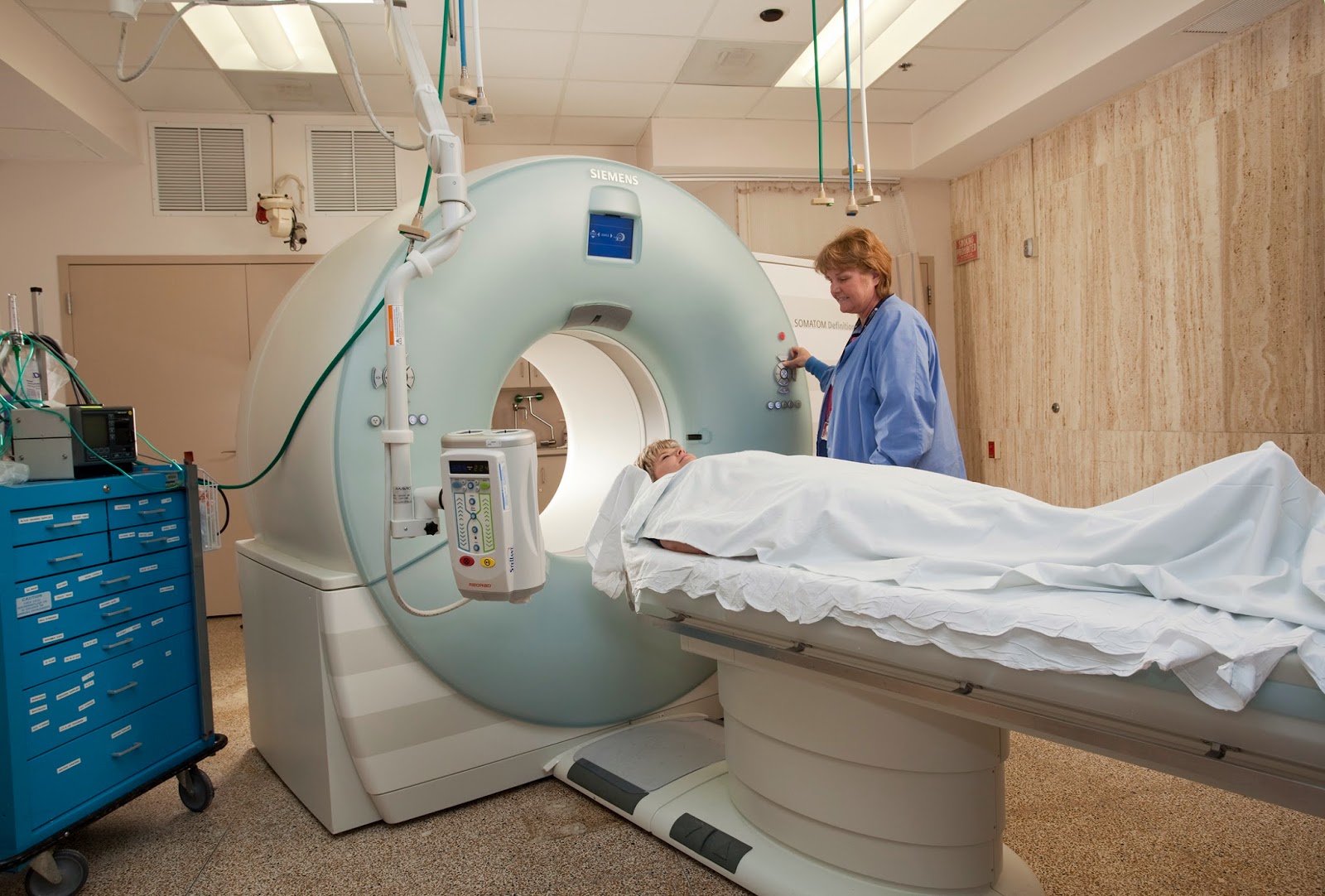
CT scans – also referred to as computerized axial tomography (CAT) – are special X-ray tests that produce cross-sectional images of the body using X-rays and a computer. CT was developed independently by a British engineer named Sir Godfrey Hounsfield and Dr. Alan Cormack and were jointly awarded the Nobel Prize in 1979. Yes, more Nobel prize winners…


In a nutshell, x-ray computed tomography:


– uses data from several X-ray images of structures inside the body and converts them into 3D pictures – especially useful for soft tissues.


– emits a series of narrow beams through the human body, producing more detail than standard single beam X-rays.


– is able to distinguish tissues inside a solid organ. A CT scan is able to illustrate organ tear and organ injury quickly and so is often used for accident victims.


– is analyzed by radiologists.


Unfortunately, unlike MRI scans, a CT scan uses X-rays and therefore are a source of ionizing radiation.


Now for the fun part (see the rules here), using CAT Scan in a sentence by the end of the day:

Serious: Hey Bob, did you know that the recorded image of a CAT Scan is called a tomogram? 

Less serious: My GP suggested that howling at the moon at night is not normal behavior and he wants to send me for a CAT scan. What? No way, I’m allergic to cats…

OK, listen to Cat Stevens to decompress and I’ll see you in the blogosphere…


Pascal Tyrrell

MiWord of the day is… Atom!

MiWord, a post on Sunday?!!! Well, I have been very busy lately and fell behind on my blog so I am now playing a little catch-up…


I was waiting in Logan airport for my flight back from a presentation in Boston – what unbelievably crazy traffic in that city – and I was texting my kids with my laptop open and my tablet next to me on the seat when I thought: I feel a little like Jimmy Neutron! I enjoyed watching that show with my kids. Lots of fun. Anyway, that idea of crazy science and the internal structure of the atom as displayed on Jimmy’s t-shirt may be the premise for a great kids show but it also led to the development of MRIWhat?!!! You say. 


MRI is an imaging technique. Maybe so, but it is particular in that it does not use any classic photographic equipment (film or lenses) or use x-rays as Roentgen did. It simply numerically measures how hydrogen nuclei absorb and release energy in response to particular frequencies. Need a refresher on the structure of an atom? See this post.


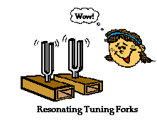
Don’t get it? OK, how about you think of this process as a crazy huge tuning fork. If you were to flick a tuning fork of a certain frequency (pitch) other tuning forks of the same frequency close by will pick up energy from the humming tuning fork and emit a sound in turn. Cool.


The nucleus of an atom can absorb energy and then relax by emitting energy in a similar way. Different atoms (or the same atom in different environments) will have different relaxation rates allowing for the identification of the composition of molecules. Ya, maybe a little complicated.


MRI measures how hydrogen nuclei absorb and release energy. Dependent on the location and the environment of the hydrogen atoms the MRI process is able to provide knowledge about the placement of hydrogen atoms in the body and in turn knowledge about the anatomy.


Now for the fun part (see the rules here), using Atom in a sentence by the end of the day:

Serious: Hey Bob, did you know that the atom is the smallest unit that defines the chemical elements and their isotopes?

Less serious: I thought that splitting atoms would produce a large explosion but when I tried using my mom’s perfume “atomizer” it just produced a fine spray and nice smell…


Ok that was a little intense for a Sunday. Watch and listen to Symphony of Science (very cool BBC production) to decompress and I’ll see you in the blogosphere…


Pascal Tyrrell

MiWord of the Day Is… Cinemaradiology!

Yes, it is Halloween today and my kids could barely contain themselves getting ready for school. I suspect today will not be very productive as they count down the minutes before heading out to terrorize my neighbors.


Anyway, how about this for a scary thought: cinemaradiology! In the late 1800’s John MacIntyre at the Gasgow Royal Infirmary experimented with producing X-ray motion pictures. What!!!? He tried exposing film by passing it between the screen of the fluoroscope and the x-ray tube and by simply filming the fluoroscopic screen. This latter method was very difficult because, as all of you budding radiologists know, the images viewed on the fluoroscope screen were dim and of poor resolution at the time.


For years researchers worked on perfecting cinemaradiology. However, during those early years of discovery they lost interest when they realized that sharper images were possible when BOTH patients and investigators were exposed together AND that excessive radiation was a bad thing – duh! 


It would only be many many years later that fluoroscope screen technology would be improved to allow for brighter and higher resolution images (and without frying the patient and everyone around!). 

Fluoroscopy is a study of moving body structures – similar to an x-ray “movie.”  A continuous x-ray beam is passed through the body part being examined, and is transmitted to a TV-like monitor so that the body part and its motion can be seen in detail. As an imaging tool, Fluoroscopy is used in many types of examinations and procedures.

To my knowledge, no actors from the cinemaradiology era ever became successful stars in Hollywood…


No need to use the MiWord of the day in a sentence today (see rules here) as I realize you are busy getting ready for Halloween and need a break!


Decompress listening to the classic song Thriller by the King of Pop Michael Jackson and I’ll see you in the blogosphere…


Pascal Tyrrell


A YSP Student Perspective: MRI and Carotid Artery Disease

Hershel Stark, MED YSP 2014 Student

Throughout the month of July, I participated in a research program with the Division of Teaching Laboratories within the Faculty of Medicine at the University of Toronto. I was assigned to work with Prof. Pascal
Tyrrell and the Department of Medical Imaging, and spent the majority of my time with the Vascular Biology Imaging Research Group (VBIRG) at Sunnybrook Research Institute. I would like to discuss my experiences, what I gained from the program, and how I can take those skills with me into the future.


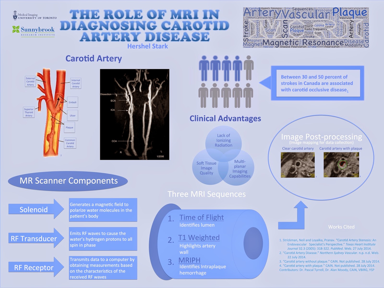
Essentially, the program was composed of presentations and shadowing opportunities in which I was introduced to various imaging modalities used in both the clinical and research fields. I primarily studied MR imaging, but was nevertheless exposed to other modalities including ultrasound and CT.  Towards the end of the program, I had two principal objectives: to present my experiences to the VBIRG group and to design an infographic for displaying. Below is a copy of my infographic:

Notwithstanding the abundance of knowledge I gained from studying the subject content, I acquired a variety of essential research skills by partaking in the program. Shadowing proficient researchers as they collected
and analyzed data provided me with a thorough insight of a researcher’s methods and techniques. The researchers that I worked with appropriately explained their individual roles on the research team, which led to my understanding of the significance of collaboration in scientific and medical research.
One last aspect of the program that I would like to address is the daily workshops that were conducted by two instructors from the Division of Teaching Laboratories, Jastaran Singh and Jabir Mohamed. Each of these brief workshops focused on an important general topic relevant to research in general, ranging from discussing common scientific practices to elaborating on literary research. I believe that the combination of skills and knowledge that I obtained from all elements of the program will be useful in my potential
research career in university.
 Lastly, I would like to take this opportunity to formally thank all of those that contributed to making the program a truly enjoyable and intellectually stimulating experience. I would like to extend my gratitude to
Dr. Alan Moody and the members of the VBIRG group for allowing me to shadow their research projects, as well as to Prof. Pascal Tyrrell and the Department of Medical Imaging at U of T for constructing the program and offering much assistance in the formation of my infographic. Finally, I’d like to thank Dr. Chris Perumalla and the Division of Teaching Laboratories in the Faculty of Medicine at U of T for formulating the research module of the Youth Summer Program, and Jastaran Singh and Jabir Mohamed for providing guidance as instructors throughout the program.
Best of luck in all of your future endeavours,
Hershel Stark

A YSP Student Perspective: Ultrasound – Not Just for the Unborn Child

Angela Lo, MED YSP 2014 Student
When you think of ultrasound, what’s the first thing you think of? Babies. All that fun stuff. Well, it turns out that ultrasound can be used not only for clinical testing, but also for research purposes. For
example, it can be used as a diagnostic tool to survey images of the body or also used as a device that monitors health conditions in research studies.
During the past three weeks, I (Angela!) have had the opportunity to participate in a research module in the medical imaging department at The University of Toronto. Through this program, I have been exposed to various imaging modalities including both MRI machines and CT scanners, but one of the modalities that interested me most was the ultrasound machine. Why? Because of its noninvasive procedures and its ability to make both 2D and 3D images in real time while still being a fraction of  he cost of an MRI.
Amazing.
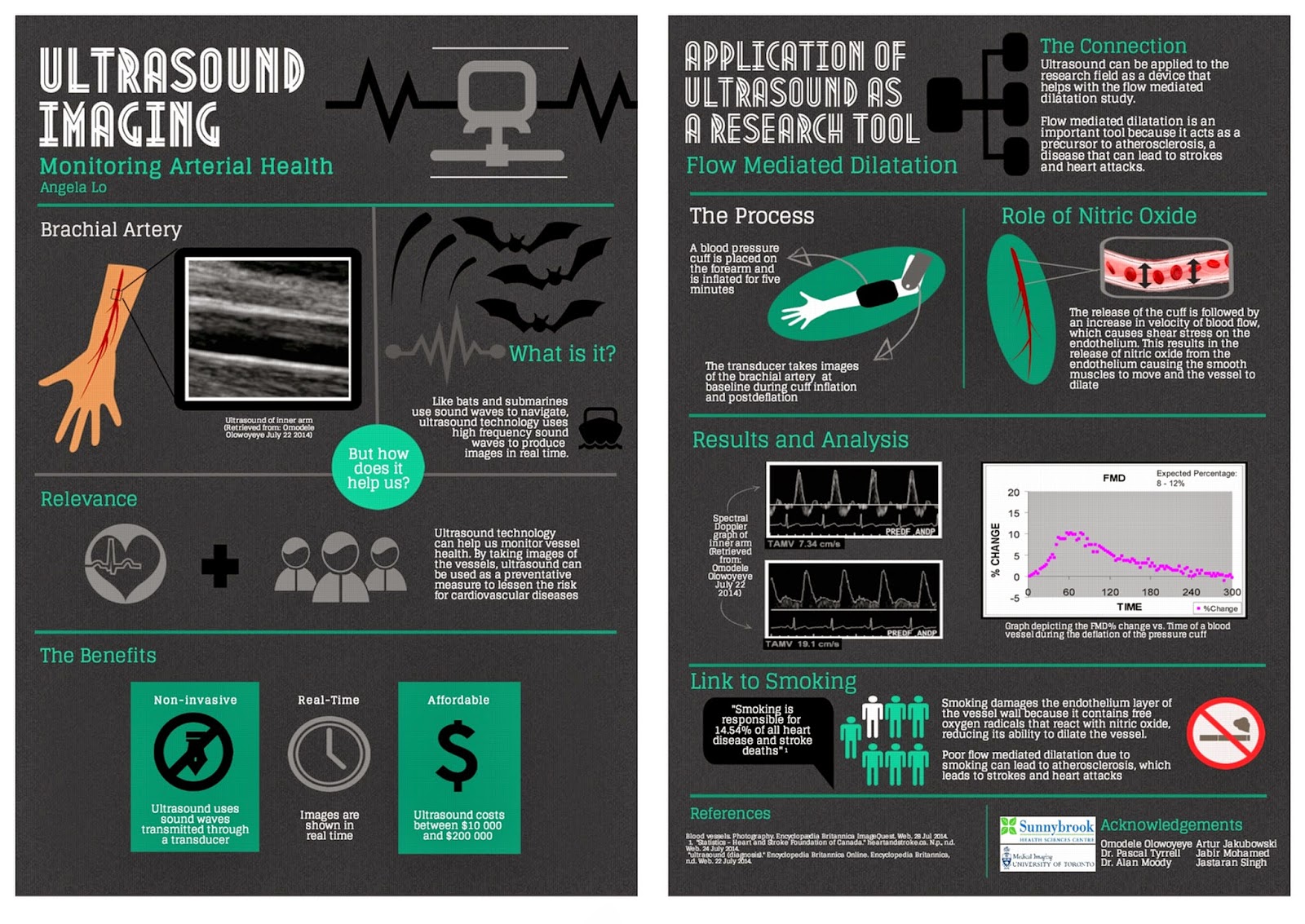
During my time in the program, I was also able to observe the various uses of the ultrasound machine and how it can be used as a research tool in the flow mediated dilatation study. Blood vessel health can be studied by having an ultrasound take images of the brachial artery to measure blood velocity and the percent change in FMD. By knowing the percent change, researchers can monitor arterial health and use it as a preventative measure.
Overall I had an amazing experience learning about all he imaging modalities and the great benefits and potential each one holds.
Happy reading,
Angela Lo

MiWord of the Day Is… Pentimenti!

What!!? Do you find it on pizza or in the middle of green olives? Well actually, it is a word of Italian origin and describes minor changes in a painting during its composition. So, similar to erasing some of your hand writing and then writing over it again with the corrected text. I guess for you younger folk it would be like pressing back-space and then re-typing! The difference of course is that there would NOT be any pentimenti as there would no trace of your previous attempt…


So what does this have to do with medical imaging? In our last Mi word of the day we talked about x-rays. Now, today we take x-rays and our ability to peer inside the human body for granted. So what else can we see with x-rays? Believe it or not x-rays can also help to reveal how a painting evolved from first brush strokes to finished product. X-ray analysis can help to describe the paint composition to the different layers that may exist in the painting.


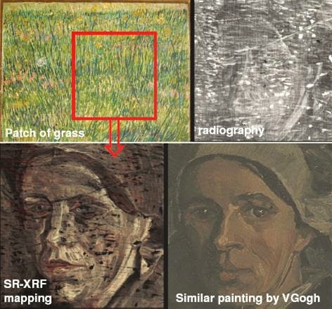
Consider, for example, “Patch of Grass” by Van Gogh seen above. It was discovered by x-ray analysis that this 1887 painting completely concealed a portrait of a woman that Van Gogh had painted over. He often did this to save money on canvases (maybe to buy Absinthe – how naughty!). In this case, in addition to Van Gogh’s pentimenti is his habit of painting over previous works. All of this adds to a type of “fingerprint” that art appraisers use to identify works of art from forgeries… Cool.

Today, we have to use “pentimenti” in a sentence (see rules here). Here are two examples to help you along:

Serious: I wonder how Van Gogh’s pentimenti differs from that of Rembrandt. Maybe I should ask the Musee du Louvre’s curator for some insight.

 
Not so serious: Yes, I would like to order a large pizza with pentimenti, double cheese, and mushrooms. No pentimenti? Alright, pepperoni works just as well…


See you in the blogosphere,


Pascal Tyrrell

Breaking Up Is Hard to Do

Last week I met with Helen, a clinical investigator program radiology resident from our department, about her research (shout out to Dr Laurent Milot’s research group). When discussing predictors and outcomes for her retrospective study it was suggested that some continuous variables be broken up into levels or categories based on given cut-points. This practice is often encountered in the world of medical research. The main reason? People in the medical community find it easier to understand results that are expressed as proportions, odds ratio, or relative risk. When working with continuous variables we end up talking about parameter estimates / beta weights and such – not as “reader friendly”. 


Unfortunately, as Neil Sedaka sang about in his famous song Breaking Up Is Hard to Do, by breaking up continuous variables you pay a stiff penalty when it comes to your ability to describe the relationship that you are interested in and the sample size requirements (see loss of power) of your study.


You are now a newly minted research scientist (need a refresher? See Pocket Protector) and are interested in discovering relationships among variables or between predictors and outcomes. The more accurate your findings the better the description of the relationships and the better the interpretation/ conclusions you can make.The bottom line is that dichotomizing/ categorizing a continuous measure will result in loss of information. Essentially, the “signal” which is the information captured by your measure will be reduced by categorization and, therefore, when you perform a statistical test that compares this signal to the “noise” or error of the model (observed differences between your patients for example) you will find yourself at a disadvantage (loss of power)David Streiner (great author and great guy!) gives a more complete explanation in one of his papers.


Now, as we see in the funny movie with Vince Vaugh and Jennifer Aniston, The Break Up, there are times when categorization may make sense. For example when the variable you are considering is not normally distributed (see Are You My Type?) or when the relationship that you are studying is not linear. We will talk about these situations in a later post.


Don’t forget: you will get further ahead if you keep your variables as continuous data whenever possible.


See you in the blogosphere,


Pascal Tyrrell

MiWord of the Day Is… X-Ray!

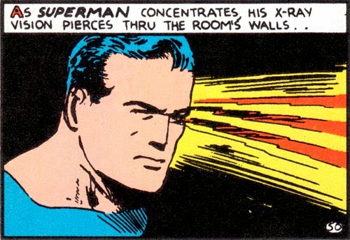
Yup! Want some of that. Not only is Superman cool but he has x-ray vision. Unbelievable. Or is it? Radiologists have the same x-ray vision but without the Spandex suit – or at least they don’t wear it to work that I am aware of.


The word of the day is x-ray. You have already successfully used “Roentgen” in casual conversation last week (don’t know what I am talking about? See Mi Word of the Day Is… Roentgen!) and today I will talk a little about what Roentgen was first in measuring and describing – x-rays.


Let’s say you are in your lab and you are working with passing electrical discharges through vacuum tubes – a typical Saturday afternoon activity with friends. As chance would have it your little sister’s barium salts paintings happen to be drying near-by and you notice a faint glow emanating from them every time you run your experiments. No matter how much you try to block any light coming from your vacuum tubes the glow persists. What? That’s odd. How’s that happening? Well my friend, you have just crossed over into the Twilight Zone (awesome old tv series) and discovered a form of electromagnetic radiation.


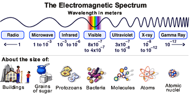
Visible light is but a very small part of the electromagnetic spectrum. Moving from visible light to longer wavelengths and lower frequencies we find infrared (keeps food warm at restaurants), microwaves (to warm your pizza pop) and radio (not the one streamed through the internet!). 


Now if you move in the opposite direction from visible light you find shorter wavelengths with higher frequencies starting with ultraviolet (what helps you get that summer tan), x-rays (word of the day), and finally gamma rays (topic for another day!). So x-rays are about the size of atoms and radio waves the size of buildings. Crazy. I think what is surprising is that with the naked eye we “see” so little and yet so much (philosophy anyone?).


So, x-rays are short wavelength, high frequency, high energy electromagnetic radiation that is able to penetrate some substances more easily than others. For example, they penetrate flesh more easily than bone, and bone more easily than lead. Thus they make it possible to see bones within flesh and a bullet embedded in bone. The ability of X rays to penetrate depends on their wavelength and on the density and thickness of the substance being scanned.


Now if you remember the rules:


1- I introduce and discuss a word.
2- You have to use the word in a sentence by the end of the day. No need to use it in the correct context – actually out of context is more fun and elicits a more entertaining response!
 
 
Today, we have to use “x-ray” in a sentence. Here are two examples to help you along:

Serious: Hey Frank, did you know the radiation you received during your chest x-ray last week was actually “soft” x-rays? Ones with shorter wavelengths and more penetrating power are used for scanning archaeological artifacts.


Less serious: Frank! Dude, I got them! My x-ray specs just came in the mail. Let’s go the beach…

See you in the blogosphere,


Pascal Tyrrell