Some R&R with UofT YSP and Elizabeth Lehner…

Elizabeth Lehner – YSP 2015


Maybe not all rest and relaxation but certainly radiology and rheumatology! Here is a great example of why collaboration between disciplines is so important in medicine. Elizabeth recently graduated from Iroquois Ridge High School and will be a new University of Toronto student this fall. See her post below. 


Great job Elizabeth!!!

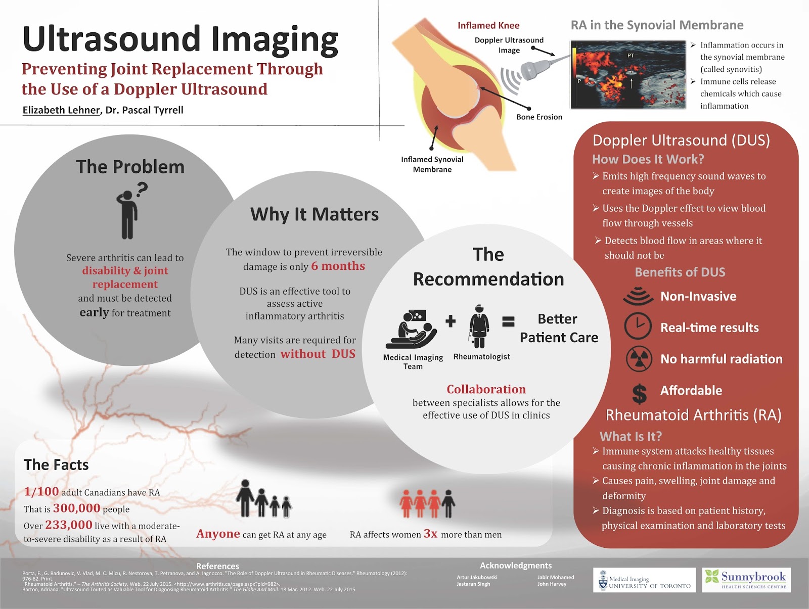
Many people are familiar with the word arthritis. This is probably because one in six Canadians aged 15 years and older report having arthritis. Rheumatoid Arthritis is a specific form of arthritis that unfortunately can lead to severe disability and joint replacement. 


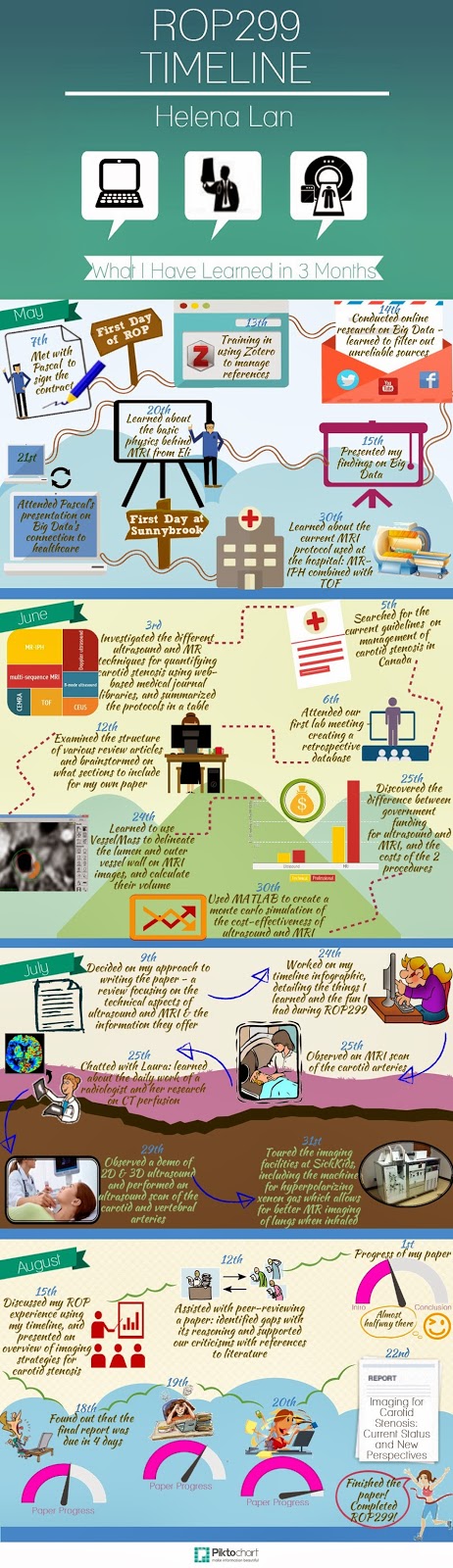
Over the past several weeks, I participated in the 2015 YSP research program with the Division of Teaching Laboratories within the Faculty of Medicine at the University of Toronto and had the opportunity to look more closely at Rheumatoid Arthritis and ways to better diagnose this debilitating disease.


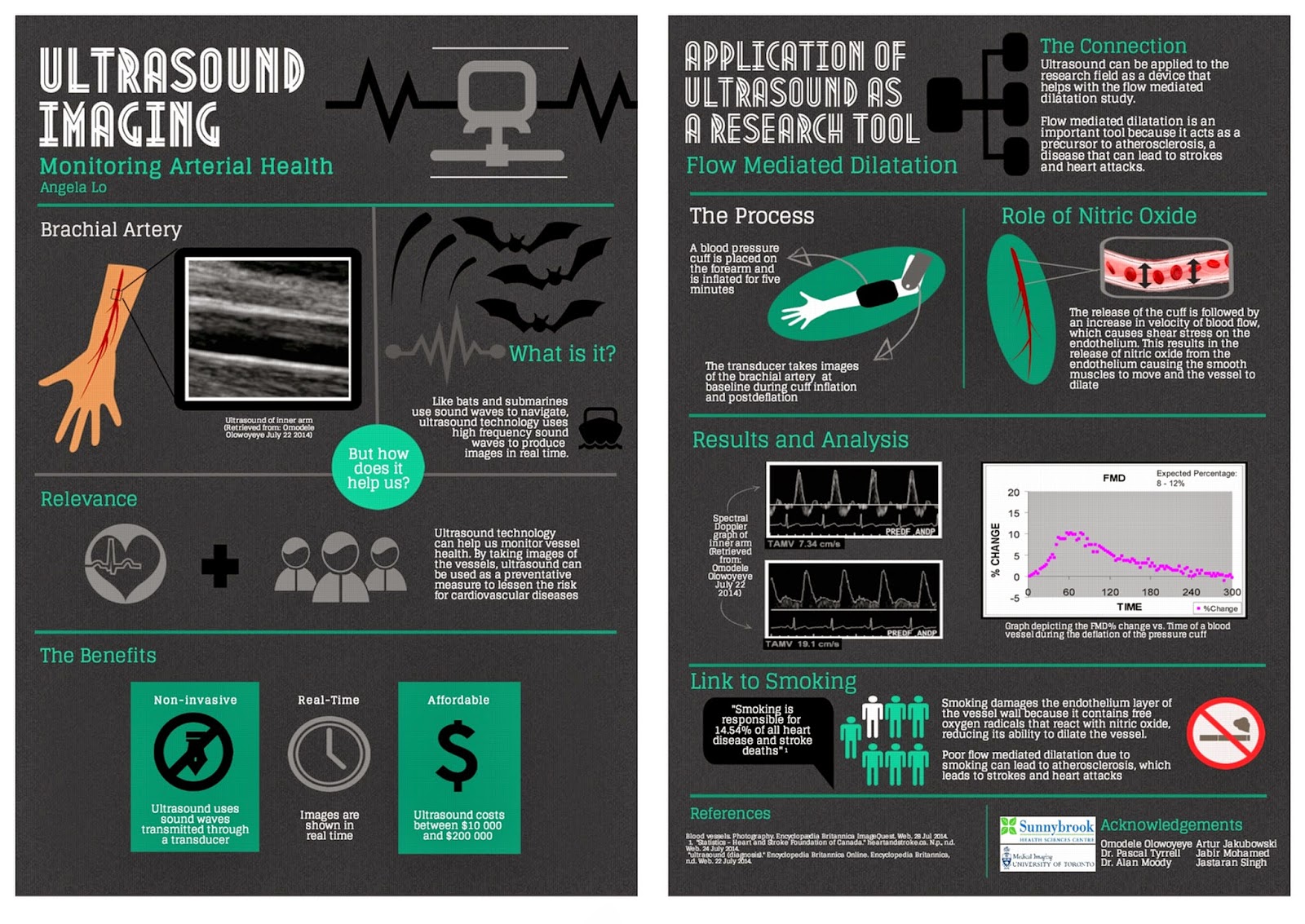
Under the supervision of Prof. Pascal Tyrrell and the Department of Medical Imaging at U of T, I was introduced to various imaging modalities including MRI machines, CT scanners and ultrasound machines. The work by Dr. Tyrrell was of particular interest given his studies on inflammation and the use of the various imaging modalities.


As part of this program I also participated in specific lab tasks including dissections and micropipetting and was exposed to clinical work such as suturing and operating an ultrasound machine. In addition, the program provided me with the opportunity to participate in daily workshops led by two instructors from the Division of Teaching Laboratories, Jastaran Singh and Jabir Mohamed. These workshops provided important overviews on a variety of topics relating to research that were very interesting.


The things I learned in this program provided me with a much better understanding of various research and medical issues that I think will be of use to me as I begin my studies at the University of Toronto this fall.
I would very much like to thank Prof. Pascal Tyrrell, Jastaran Singh and Jabir Mohamed for allowing me to be exposed to the various projects and for answering the many questions that I had during the program. Thank you!

 
Elizabeth Lehner

The story behind Connectory…

So, you are reading our blog thinking Pascal is a nut – that much is clear – but what of all the students plugged into his group? Are they nuts too?


Well maybe, but today I am going to talk to you about the group of four (not the group of seven) who started small and grew to be Connectory. John, Maria, Natasha, and Roger met in a graduate course at the University of Toronto and decided to work together on a project about innovation. That’s when they met me, joined “the program”, and got busy! Starting any endeavour from scratch is no easy task. All four had never met before, all came from very different academic backgrounds, and though their initial project was for “credit” the rest was on their own time.


There were some rough times at first but with perseverance comes success and Connectory was born and is just finishing up its first project as a new start-up business. Wow! 


Essentially Connectory is a data management solutions software development consulting group that operates in the healthcare space. Check out their webpage here.


Ok, so what? Well this post is not only to congratulate these four on a job well done but also to encourage you to do the same. One thing is for sure: if you don’t try you will not succeed – ever. My programs are all about learning, trying new stuff, benefiting from your successes as well as your failures, and wait for it… giving back. Yup as Uncle Ben said in Spiderman: “With great power comes great responsibility“. 


Just wanted to share a good story from our group with you today.


Listen to Bulletproof by La Roux to get pumped and…


… I’ll see you in the blogosphere!


Pascal Tyrrell

Happy Late New Year 2015!!!

The Moody Tree


Ok, so I may have taken a longer break than I should have. Where was I you ask? I was enjoying some R&R with my family. My kids are at great ages – 15, 11, and 6. Then, of course, when I got back to my desk – whammo! The deluge of work. This morning, as I sat on the GoTrain on the way into Toronto, I thought of you and happily sat down to write my first post of 2015.


First, a thank you for your readership. We are soon approaching our first anniversary (next month) and my programs (MiVIP and MiB) and this blog are chugging along famously… all because of you!


Chicago Bean

Next, a funny story to explain the picture above. When I attended the RSNA last December (see my post here on this event) I brought along my old film camera for fun as I enjoy photography and decided to reminisce a little. To your right is a picture of the Chicago skyline reflected on the “Bean“. See me?


I hadn’t developed film in so long that I almost ruined it – in my laundry room between all of my family’s clothes, the ironing board, buckets, detergents… Anyway, I also had with me my trusted digital for snaps and one evening I was invited to a function at my boss’ hotel and he said to me:”Let me know what you think of the Christmas lights on the trees in front the hotel on your way in”. Alan Moody is an uber-radiologist, the chair of our department, and loves imaging the carotid arteries. As our minds often operate on the same wave lengths, I took the picture and voila – the Moody Tree was born!


There is no end to the fun we have here in the Department of Medical Imaging


Even though this is not a “MiWORD” post how about you wish a belated Happy New Year to someone you have not been in touch with yet? Send them a quick text or better yet, send them the link to this post and tell them to visit the Moody Tree next time they are in Chicago during the holidays…


See you in the blogosphere,


Pascal


MiWord of the day is… Atom!

MiWord, a post on Sunday?!!! Well, I have been very busy lately and fell behind on my blog so I am now playing a little catch-up…


I was waiting in Logan airport for my flight back from a presentation in Boston – what unbelievably crazy traffic in that city – and I was texting my kids with my laptop open and my tablet next to me on the seat when I thought: I feel a little like Jimmy Neutron! I enjoyed watching that show with my kids. Lots of fun. Anyway, that idea of crazy science and the internal structure of the atom as displayed on Jimmy’s t-shirt may be the premise for a great kids show but it also led to the development of MRIWhat?!!! You say. 


MRI is an imaging technique. Maybe so, but it is particular in that it does not use any classic photographic equipment (film or lenses) or use x-rays as Roentgen did. It simply numerically measures how hydrogen nuclei absorb and release energy in response to particular frequencies. Need a refresher on the structure of an atom? See this post.


Don’t get it? OK, how about you think of this process as a crazy huge tuning fork. If you were to flick a tuning fork of a certain frequency (pitch) other tuning forks of the same frequency close by will pick up energy from the humming tuning fork and emit a sound in turn. Cool.


The nucleus of an atom can absorb energy and then relax by emitting energy in a similar way. Different atoms (or the same atom in different environments) will have different relaxation rates allowing for the identification of the composition of molecules. Ya, maybe a little complicated.


MRI measures how hydrogen nuclei absorb and release energy. Dependent on the location and the environment of the hydrogen atoms the MRI process is able to provide knowledge about the placement of hydrogen atoms in the body and in turn knowledge about the anatomy.


Now for the fun part (see the rules here), using Atom in a sentence by the end of the day:

Serious: Hey Bob, did you know that the atom is the smallest unit that defines the chemical elements and their isotopes?

Less serious: I thought that splitting atoms would produce a large explosion but when I tried using my mom’s perfume “atomizer” it just produced a fine spray and nice smell…


Ok that was a little intense for a Sunday. Watch and listen to Symphony of Science (very cool BBC production) to decompress and I’ll see you in the blogosphere…


Pascal Tyrrell

MiWord of the Day Is… Cuckoo!

One of my favorite more serious films is One Flew Over the Cuckoo’s Nest. What does Jack Nicholson’s portrayal of a bad guy hoping for easy served time in a mental institution have to do with medical imaging? Well it all starts with the lobotomy. Not to spoil the story, suffice it to say that the movie broaches the topic of lobotomies and how ridiculous they were. Lobotomy was a form of neurosurgery that involved damaging the prefrontal cortex in order to “calm” certain mentally ill patients. Needless to say the procedure was controversial from the beginning (1935 to the early 1970’s) but the author of the discovery, Egas Moniz, was awarded the Nobel Prize in 1949. Maybe not the most sound of decisions by the committee. However, for the time, it was considered progress in a very challenging area of medicine – mental illness. 


OK, medical imaging? Well as it turns out Moniz (do not confuse with St-Moriz, ahhh skiing…) is also known for developing cerebral angiography – a technique allowing the visualization of blood vessels in and around the brain. 


Moniz was interested in finding a non-toxic substance that would be eliminated from the body, but would not be diluted by the flow of blood before the x-ray could be taken. Another requirement is that the substance could not cause an emboli or clot as this would be a bad thing. Moniz played with salts of iodine and bromine and settled on iodine because of its greater radiographic density. And voila, birth of iodinated radiocontrast agents still in use today. Cool.


Supposedly it took him 9 patients to perfect his angiogram technique. Don’t ask about the first 8…

Moral of the story is: lobotomy bad and cerebral angiography good.


Now for the fun part (see the rules here), using Cuckoo in a sentence by the end of the day:

 
Serious: Hey Bob, when I was visiting my aunt in Australia I spied a little bronze cuckoo in her backyard! This could be my “big year“…


Less serious: Someone won a Nobel Prize for developing the lobotomy? Are you cuckoo?
 
 
Listen to Los Lobos (not short for lobotomy but “the wolves” in Spanish) singing La Bamba to decompress and…
 
… I’ll see you in the blogosphere,
 
 
Pascal Tyrrell

Starting a new research project? Think Zorro!

Well, OK maybe think Zotero. The Mask of Zorro was such a great movie I could not resist. Having said that, when starting a new research project it may be helpful for you to think of yourself as Zorro. It may give you that extra zip required to get you through the inevitable research project doldrums…


So what is this Zotero thing anyway? Well Zotero is an open source reference management software that can act as your personal research assistant – helping you to organize and cite the numerous articles that you will be reviewing.


I was talking to Ori the other day – who is in the Radiation Therapy program at the Michener Institute – and he is in the process of planning a research project. As it turns out he has been a member of the MiVIP family since the beginning so he is well aware of my earlier posts that will help him along:


1- Thoughts on how to become a researcher


2- What is in a research question?


3- What makes up a good research question with the F.I.N.E.R. series.


Now how about the reference management software thing? Well, I give you an easy, fun, and instructional e-learning module to help you along. Our group has just finished our first kick at the can (so to speak) and so I invite you to have a look. Here is the link:


MiEducation Zotero e-learning module


Tell us what you think by posting comments and suggestions to this post.


Maybe listen to Ylvis in What Does the Zorro Say? while you go through the module. Fox in spanish is zorro…

 
… and I’ll see you in the blogosphere.
 
 
Pascal Tyrrell


MiWord of the Day Is… Ionizing!

So what the heck is ionizing radiation? Well consider the following conundrum about x-rays. They can:


1- cause cancer
2- be used to detect cancer.
3- be used to treat cancer.


Whaaat? How does that work?  We use the term ionizing  when the radiation has sufficient energy to detach electrons from molecules causing them to become chemically reactive ions


The name atom means “indivisable” and are incredibly small. They are made up of protons, neutrons, and electrons with about 99.9% of its mass concentrated in the nucleus that holds a positive charge. A surrounding negatively charged cloud of electrons makes up the difference and the atom stays together due to the attraction between the two. 


OK, so here is the rub: if an atom gains or loses an electron it becomes an ion and generally results in a very chemically reactive substance. This process to produce an ion can be achieved by many ways but one of the most important is electromagnetic radiation (we’ve talked about this already here). Radioactive materials such as radium emit ionizing radiation as does x-ray tubes. There is even such a thing as cosmic radiation (Yup, we talked about that here!). 


Now x-rays produce photons which are the same particles that make up visible light but at a much shorter wavelength and higher energy. When they penetrate through a solid object they will most often simply pass through. However, if they pass by close enough to an electron they can transfer their energy and in the process knock it out orbit producing an ion. Also, the more dense the object the more often the photons are blocked from travelling through resulting in a differential effect on a film or sensor placed on the opposite side. This is how we are able to see inside the body using x-rays.


The problem about ionizing radiation is that the resulting chemically reactive ions can result in DNA damage. Often the cell can repair itself resulting in no permanent damage. Other times, however, permanent damage occurs and can result in cell death (a good thing if they are cancerous cells) or DNA mutations that can in turn lead to the promotion of cancer – bummer.


Now on to using ionizing in a sentence today (not sure about the rules? See here):
Serious example – Bob, don’t stand too close to the x-ray machine. You wouldn’t want to be exposed to ionizing radiation that could damage the DNA in your cells…


Less serious – You wouldn’t believe what happened to me at work today! I was at the photocopy machine getting ready to change the toner cartridge and Bill from sales said:”Let me do that for you, Honey”. He is so ionizing or patronizing or whatever. He makes me mad… 


Listen to Just Because by Raygun to get ready for the weekend and I’ll see you back in the blogosphere soon.


Pascal Tyrrell

MiWord of the Day Is Something to “Bragg” About…


OK who hasn’t made rock candy as a kid? No? Give it a try. Maybe you have a little brother or sister you can impress. All you need is a super saturated solution of sugar, a surface for crystal nucleation (string), and lots of time…


Now what if you were to apply this technique to obtain crystals of DNA? I don’t suggest that you eat it as a treat but you could possibly try X-ray crystallography.


The challenge is that DNA, unlike proteins, is an exceedingly large molecule which does not lend itself to crystallisation. The result is a highly viscous suspension of spiderweb-like filaments. However, it is this very suspension that the DNA molecules were deduced to be neatly aligned alongside one another by studying the X-ray diffraction patterns. This initial challenge was successfully overcome by Rosalind Franklin. Her hard work then laid the ground work for Watson and Crick to piece together the puzzle of DNA structure (winning the 1962 Nobel Prize along with Wilkins).


Now the x-ray crystallography imaging technique is no pic-nic! It was first described by the Australian father-and-son duo William Henry Bragg and William Lawrence Bragg. Essentially x-rays are projected onto a crystalline solid and when analyzing the diffraction patterns it is possible to determine how its molecular atoms are positioned in relation to one another. This is due to x-rays having very short wave-lengths (see x-rays in the blog) and the mathematical analysis of predictable diffraction from the three dimensional structure of the crystal. It was Lawrence Bragg who developed the equation to describe this diffraction and is now known as Bragg’s Law. He and his father won the Nobel Prize for this work in 1915. 


I used to listen to New Order back in the day and they are still singing (and dancing?) with a more recent release of Crystal by New Order. So, listen to the song while making rock candy and maybe you too will come up with a brilliant idea worthy of a Nobel Prize by the time the crystals are big enough to eat.


Now if you remember the rules:


1- I introduce and discuss a word.
2- You have to use the word in a sentence by the end of the day. No need to use it in the correct context – actually out of context is more fun and elicits a more entertaining response!


Today, we have to use “Bragg” in a sentence. Here are two examples to help you along:

Serious: “Hey Bob, did you know that if you used a saturated sodium salt solution of DNA instead of sugar to produce crystals we could then do some x-ray crystallography for fun and apply Bragg’s law to determine the molecular structure…”
 
Less serious: “Bob, I don’t want to Bragg but my crystals are way bigger than yours…”


See you in the blogosphere,


Pascal Tyrrell

Helena Lan: Summer 2014 ROP

Helena Lan Summer 2014 ROP

What is research like? If you had asked me this
question several months ago, I would have answered, “You wear a lab coat and
goggles while mixing chemicals or observing organisms. Hopefully something
interesting will happen, so that you get to publish your findings!” Well, after
participating in the Research Opportunity Program (ROP) at the University of
Toronto, I discovered that medical imaging research is more than just
pipetting, and is all the more exciting!

 
So what kind of research is conducted in the medical imaging world? For my ROP, the objective of my project was to evaluate the roles of the non-invasive imaging modalities for diagnosing carotid stenosis. Hence, I engaged in online literature research of the various imaging techniques for assessing this disease. In this process, I also learned to use Zotero to manage all my references, which provides an easy way to generate a bibliography (when the software doesn’t crash every time you open it). After gathering all the pertinent information, I then put together a review article suggesting how a change in the current imaging approach could potentially improve clinical outcome. Who knew a report could be compiled without doing the lab grunt work?     
 
Wait, so this is all a radiologist does? Sitting in front of a computer and typing all day? Of course not! During our time at Sunnybrook Hospital, we got the chance to chat with a radiologist and discovered that she could decide whether patients should be released after taking a look at their diagnostic images. Pretty powerful, eh? That’s not all. We also found out how radiologists identified any abnormalities in patients, as we had the opportunity to work with the VesselMass software which allowed for the delineation of the lumen and vessel wall of arteries on MRI images. Oh, and did I mention we observed an MRI and an ultrasound examination of the carotid arteries, and even got to perform an ultrasound scan ourselves. Super cool!
 
Still craving for more of my ROP experience? Check out my timeline infographic! You will find all the things I learned and all the fun I had there. Last but not least, I’d like to shout out a big THANK YOU to Prof. Pascal Tyrrell and Dr. Eli Lechtman, who guided us every step of the
way. Also, I’m very grateful to Dr. Alan Moody for including us in his research program at Sunnybrook, as well as other members of the VBIRG group who gave us the chance to
participate in various activities. My summer would not have been this fun and meaningful without all of your help!
 
Have fun researching,


Helena Lan
 

A YSP Student Perspective: Ultrasound – Not Just for the Unborn Child

Angela Lo, MED YSP 2014 Student
When you think of ultrasound, what’s the first thing you think of? Babies. All that fun stuff. Well, it turns out that ultrasound can be used not only for clinical testing, but also for research purposes. For
example, it can be used as a diagnostic tool to survey images of the body or also used as a device that monitors health conditions in research studies.
During the past three weeks, I (Angela!) have had the opportunity to participate in a research module in the medical imaging department at The University of Toronto. Through this program, I have been exposed to various imaging modalities including both MRI machines and CT scanners, but one of the modalities that interested me most was the ultrasound machine. Why? Because of its noninvasive procedures and its ability to make both 2D and 3D images in real time while still being a fraction of  he cost of an MRI.
Amazing.
During my time in the program, I was also able to observe the various uses of the ultrasound machine and how it can be used as a research tool in the flow mediated dilatation study. Blood vessel health can be studied by having an ultrasound take images of the brachial artery to measure blood velocity and the percent change in FMD. By knowing the percent change, researchers can monitor arterial health and use it as a preventative measure.
Overall I had an amazing experience learning about all he imaging modalities and the great benefits and potential each one holds.
Happy reading,
Angela Lo