Research Opportunity Program: Cost Effectiveness and Imaging Carotid Stenosis

ROP Research Forum March 5th, 2015 – Sylvia Urbanik

Sylvia is a second year student studying cell and molecular biology. She is currently finishing up her Research Opportunity Program that spanned the fall and winter semesters, and also recently represented us at the March 5th ROP research forum along with her partner and predecessors.

 
Her project dealt with cost-effectiveness analysis techniques for diagnostic imaging modalities, with a specific focus on carotid artery stenosis. She explored the different types of analyses used in assessing cost effectiveness. She examined how factors such as sensitivity and specificity of diagnostic tests (imaging modalities in this case) can affect the analysis, and conducted literature searches in order to find these variables in order to incorporate them into a cost effectiveness model.​

Well done, Sylvia!

Stay tuned, next Kevin Chen will be modeling with TreeAge…

See you in the blogosphere,

Pascal Tyrrell

Research Opportunity Program: Imaging Carotid Stenosis

ROP Research Forum March 5th, 2015 – Helena Lan

Helena is a second-year student pursuing a specialist in Pharmacology and Toxicology at the University of Toronto. Participating in ROP299 this past summer (see here) has opened many doors for her. She is currently assisting Pascal on a systematic review on research methodology and biostatistics in medical imaging, working as a research assistant for a medical education study, participating in the research abroad program at Karolinska Institutet this summer… and recently represented us at the March 5th, 2015 ROP research forum!

Helena examined the technical aspects and information provided by two major imaging modalities, MRI and ultrasound, for diagnosing carotid stenosis. She suggests that MRI holds great promise to serve as a cost effective test for carotid stenosis as well as a tool for assessing vessel health and plaque composition that would provide important information for patient management decisions.


Stay tuned, next Sylvia Urbanik will be talking about cost effectiveness…

See you in the blogosphere,

Pascal Tyrrell

Research Opportunity Program: What Is Carotid Stenosis and How Are We Treating Affected Patients?

ROP Research Forum March 5th, 2015 – Alana Man

Alana Man is a second year University of Toronto student pursuing a specialist in Bioinformatics and Computational Biology and a major in Immunology. She was a Research Opportunity student last summer with MiVIP (see here) and recently represented us at the March 5th, 2015 ROP research forum.

Alana’s project focused on care for patients with carotid stenosis in Toronto, Ontario, Canada. She looked at the different factors contributing to access to diagnostic testing such as procedure costs, wait times, and the differences between available imaging modalities and explores whether MRI could be the diagnostic test of choice over DUS for people at increased risk of carotid stenosis.

Well done, Alana!


Stay tuned, next Helena Lan will be comparing MRI and DUS…


See you in the blogosphere,


Pascal Tyrrell

MiWord of the Day Is… PET scan!


Peanuts. What a great story. The most popular and influential comic strip in history. Snoopy was my first stuffed animal growing up. He still lives with my parents. So what is a PET scan anyway? I don’t recall ever seeing the picture above in any of the Peanuts cartoon strips.


Positron emission tomography (PET) is somewhat of a special medical imaging modality in that it brings together two different technologies from different times. Let me explain. Back in the early 1930s, George Hevesy was a young Hungarian physicist who developed biologically safe and useful radioactive tracers that could be ingested or incorporated into the body in some way.  Physicians would then manually locate where these radioactive tracers had gone in the body by using a Geiger counter at first and then later using special cameras (Kuhl‘s photoscan) to produce a crude emission image. 


So, how do we get cool pictures like these ones?  Well we would have to wait another 25 years after the development of radioactive tracers by Hevesy for the start of construction of instruments able to not only detect these radioactive sources in the body but to produce tomographic pictures.


It won’t be until the mid 1970s that PET – as we know it today – would be born. Essentially, a patient receives a emissions scan (PET) and a CT (we talked about that here) or MRI (we talked about that here) scan at the same time. The two scans are then merged together thanks to highly specialized computers (see the pictures in the middle frames). Voila! PET. 


PET is both a medical and research tool. Most often used in clinical oncology (medical imaging of tumors and the search for metastases), it is also important in clinical diagnosis of certain diffuse brain diseases such Alzheimer’s disease and other types of dementia.


Relax your brain a little listening to Radioactive by Imagine Dragons and don’t forget the fun part (see the rules here), using PET scan in a sentence by the end of the day:

Serious: Hey Bob, did you know that much of the success of the PET scan is due to the development of the radiopharmaceutical FDG (deoxyglucose) that lead the way to the characterization of Parkinson’s and Huntington’s disease?

Less serious: I can’t believe they developed yet another PET scan. Wasn’t the CAT scan enough?

See you in the blogosphere,

Pascal Tyrrell


Happy Late New Year 2015!!!

The Moody Tree


Ok, so I may have taken a longer break than I should have. Where was I you ask? I was enjoying some R&R with my family. My kids are at great ages – 15, 11, and 6. Then, of course, when I got back to my desk – whammo! The deluge of work. This morning, as I sat on the GoTrain on the way into Toronto, I thought of you and happily sat down to write my first post of 2015.


First, a thank you for your readership. We are soon approaching our first anniversary (next month) and my programs (MiVIP and MiB) and this blog are chugging along famously… all because of you!


Chicago Bean

Next, a funny story to explain the picture above. When I attended the RSNA last December (see my post here on this event) I brought along my old film camera for fun as I enjoy photography and decided to reminisce a little. To your right is a picture of the Chicago skyline reflected on the “Bean“. See me?


I hadn’t developed film in so long that I almost ruined it – in my laundry room between all of my family’s clothes, the ironing board, buckets, detergents… Anyway, I also had with me my trusted digital for snaps and one evening I was invited to a function at my boss’ hotel and he said to me:”Let me know what you think of the Christmas lights on the trees in front the hotel on your way in”. Alan Moody is an uber-radiologist, the chair of our department, and loves imaging the carotid arteries. As our minds often operate on the same wave lengths, I took the picture and voila – the Moody Tree was born!


There is no end to the fun we have here in the Department of Medical Imaging


Even though this is not a “MiWORD” post how about you wish a belated Happy New Year to someone you have not been in touch with yet? Send them a quick text or better yet, send them the link to this post and tell them to visit the Moody Tree next time they are in Chicago during the holidays…


See you in the blogosphere,


Pascal


MiWord of the day is… Atom!

MiWord, a post on Sunday?!!! Well, I have been very busy lately and fell behind on my blog so I am now playing a little catch-up…


I was waiting in Logan airport for my flight back from a presentation in Boston – what unbelievably crazy traffic in that city – and I was texting my kids with my laptop open and my tablet next to me on the seat when I thought: I feel a little like Jimmy Neutron! I enjoyed watching that show with my kids. Lots of fun. Anyway, that idea of crazy science and the internal structure of the atom as displayed on Jimmy’s t-shirt may be the premise for a great kids show but it also led to the development of MRIWhat?!!! You say. 


MRI is an imaging technique. Maybe so, but it is particular in that it does not use any classic photographic equipment (film or lenses) or use x-rays as Roentgen did. It simply numerically measures how hydrogen nuclei absorb and release energy in response to particular frequencies. Need a refresher on the structure of an atom? See this post.


Don’t get it? OK, how about you think of this process as a crazy huge tuning fork. If you were to flick a tuning fork of a certain frequency (pitch) other tuning forks of the same frequency close by will pick up energy from the humming tuning fork and emit a sound in turn. Cool.


The nucleus of an atom can absorb energy and then relax by emitting energy in a similar way. Different atoms (or the same atom in different environments) will have different relaxation rates allowing for the identification of the composition of molecules. Ya, maybe a little complicated.


MRI measures how hydrogen nuclei absorb and release energy. Dependent on the location and the environment of the hydrogen atoms the MRI process is able to provide knowledge about the placement of hydrogen atoms in the body and in turn knowledge about the anatomy.


Now for the fun part (see the rules here), using Atom in a sentence by the end of the day:

Serious: Hey Bob, did you know that the atom is the smallest unit that defines the chemical elements and their isotopes?

Less serious: I thought that splitting atoms would produce a large explosion but when I tried using my mom’s perfume “atomizer” it just produced a fine spray and nice smell…


Ok that was a little intense for a Sunday. Watch and listen to Symphony of Science (very cool BBC production) to decompress and I’ll see you in the blogosphere…


Pascal Tyrrell

A Crash Course in Medical Imaging

Oddly enough, there’s been a surprising lack of content about medical imaging on a blog with medical imaging in its title. So in order to fill that void, I’ll be providing a brief history on the development of the clinical technique used to visualize the human body.

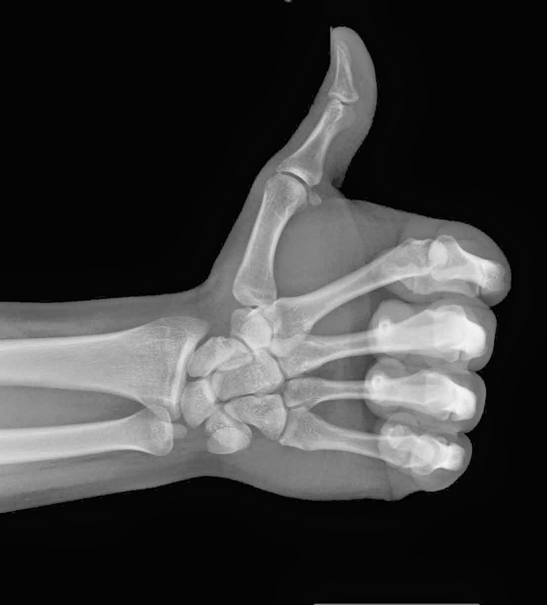
The advent of medical imaging dates all the way back to 1895, following the discovery of X-rays by the German physicist, Wilhelm Conrad Roentgen. The first X-ray picture was then produced, detailing the skeletal composition of his wife’s left hand. However, the actual quality of this imaging process was still very primitive, only allowing for the visualization of bones or foreign objects.

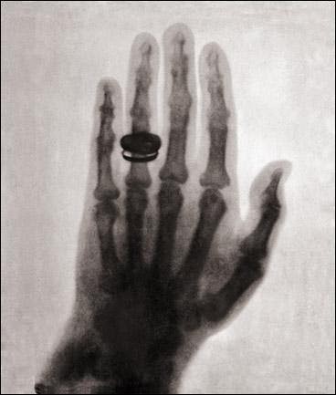
    Much to Dr. Roentgen’s pleasure, Mrs. Roentgen
    had not discarded her wedding ring
    It was not until the 1920’s that radiologists would develop a more effective method of visualization. This process, known as fluoroscopy, involved either an oral or vascular injection of a radio-opaque contrast agent, which would travel through the patient’s gastrointestinal track. Radiologists could then take films tracking the agent, allowing them to view blood vessels and digestive tracks alike.

      By the 1950’s, imaging procedures progressed towards nuclear medicine, involving radioactive compounds. These compounds were administered to patients because they could be absorbed by cellular clusters being invaded by tumours. As compounds decayed and emitted gamma rays, the recorded radiation could then be detected by gamma cameras, signalling the location of any cancerous developments. 
          The 1970’s were a period of rapid advancement for the field, as a number of modern imaging techniques were developed for clinical practice such as: 

            • Ultrasound – Uses sound waves that are able to penetrate cellular tissue. Once they reflect off the body’s internal organs, the vibrations generate an electrical pulse which can then be reconstructed into an image. 
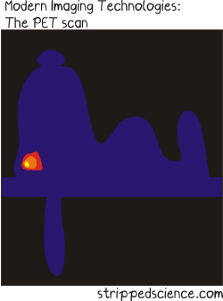
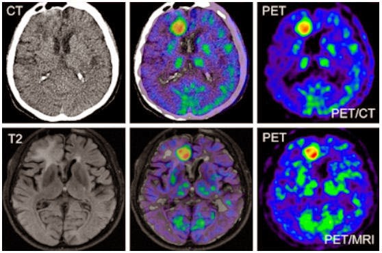
            • PET-CT Scan – Positron emission tomography (PET) uses compounds that emit positrons when they decay rather than gamma rays. It is now combined with a computed tomography (CT) device to generate a high-resolution image displaying sectioned layers of the scanned area. 
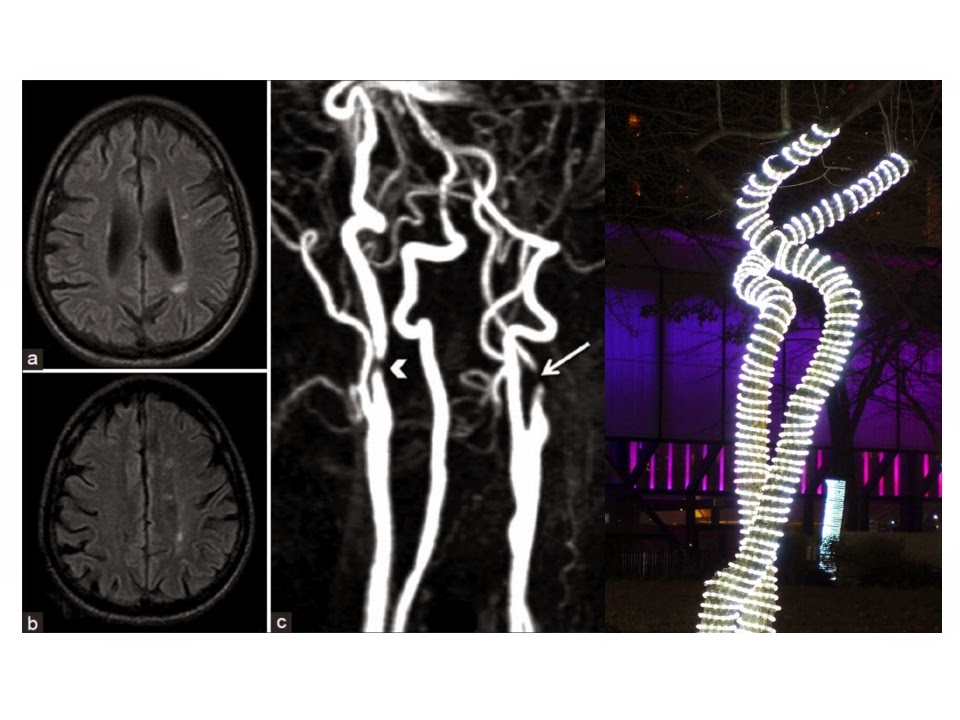
            • MRI – A Magnetic Resonance Imaging scanner runs a strong magnetic field through the body, aligning hydrogen protons. As the protons return to their original position in the atom, they generate radio waves, which are then picked up by the scanner and used to create an image based on signal strength. 

            Fast-forward to present day and over 70 million CT scans, 30 million MRI scans and 2 billion X-rays have been performed worldwide! The field of medical imaging is still growing by the day, with ongoing research leading to new developments.

              Thanks for reading,

                Brandon Teteruck