Another Reason Why a Brain Boo-Boo Is Bad.

Rostam Rashidkhani – YSP 2015


Rostam Rashidkhani is a grade 12 International Baccalaureate student at the Toronto French School and he was a Meds – Youth Summer Program student with me this summer.

Rostam is intrigued by the sciences and enjoys biology, chemistry, and physics in school. He has participated in a number of University of Toronto summer programs and is looking forward to University life!


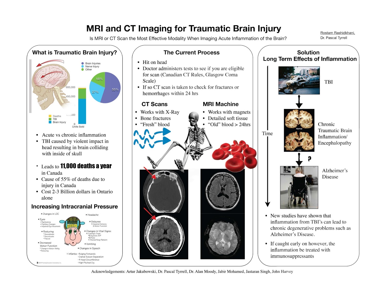
This summer Rostam looked at what causes brain problems after traumatic brain injury and how best to detect these changes with MRI. Recent brain imaging studies, including those in former professional football players, indicate that persistent brain inflammation after a single moderate head injury or repeated milder traumatic brain injury may be very common, may contribute to cognitive problems. More importantly, the chronic brain inflammation related to traumatic brain injury may be treatable. Looking for chronic traumatic brain inflammation with followup MR may be a way to reduce cognitive impairment.

Well done, Rostam!


Enjoy the read and…


… see you in the blogosphere,

Pascal Tyrrell

MiWord of the Day Is… PET scan!


Peanuts. What a great story. The most popular and influential comic strip in history. Snoopy was my first stuffed animal growing up. He still lives with my parents. So what is a PET scan anyway? I don’t recall ever seeing the picture above in any of the Peanuts cartoon strips.


Positron emission tomography (PET) is somewhat of a special medical imaging modality in that it brings together two different technologies from different times. Let me explain. Back in the early 1930s, George Hevesy was a young Hungarian physicist who developed biologically safe and useful radioactive tracers that could be ingested or incorporated into the body in some way.  Physicians would then manually locate where these radioactive tracers had gone in the body by using a Geiger counter at first and then later using special cameras (Kuhl‘s photoscan) to produce a crude emission image. 


So, how do we get cool pictures like these ones?  Well we would have to wait another 25 years after the development of radioactive tracers by Hevesy for the start of construction of instruments able to not only detect these radioactive sources in the body but to produce tomographic pictures.


It won’t be until the mid 1970s that PET – as we know it today – would be born. Essentially, a patient receives a emissions scan (PET) and a CT (we talked about that here) or MRI (we talked about that here) scan at the same time. The two scans are then merged together thanks to highly specialized computers (see the pictures in the middle frames). Voila! PET. 


PET is both a medical and research tool. Most often used in clinical oncology (medical imaging of tumors and the search for metastases), it is also important in clinical diagnosis of certain diffuse brain diseases such Alzheimer’s disease and other types of dementia.


Relax your brain a little listening to Radioactive by Imagine Dragons and don’t forget the fun part (see the rules here), using PET scan in a sentence by the end of the day:

Serious: Hey Bob, did you know that much of the success of the PET scan is due to the development of the radiopharmaceutical FDG (deoxyglucose) that lead the way to the characterization of Parkinson’s and Huntington’s disease?

Less serious: I can’t believe they developed yet another PET scan. Wasn’t the CAT scan enough?

See you in the blogosphere,

Pascal Tyrrell