MiWord of the day is… Atom!

MiWord, a post on Sunday?!!! Well, I have been very busy lately and fell behind on my blog so I am now playing a little catch-up…


I was waiting in Logan airport for my flight back from a presentation in Boston – what unbelievably crazy traffic in that city – and I was texting my kids with my laptop open and my tablet next to me on the seat when I thought: I feel a little like Jimmy Neutron! I enjoyed watching that show with my kids. Lots of fun. Anyway, that idea of crazy science and the internal structure of the atom as displayed on Jimmy’s t-shirt may be the premise for a great kids show but it also led to the development of MRIWhat?!!! You say. 


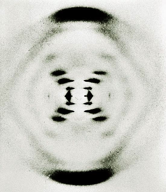
MRI is an imaging technique. Maybe so, but it is particular in that it does not use any classic photographic equipment (film or lenses) or use x-rays as Roentgen did. It simply numerically measures how hydrogen nuclei absorb and release energy in response to particular frequencies. Need a refresher on the structure of an atom? See this post.


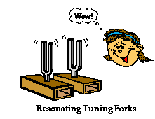
Don’t get it? OK, how about you think of this process as a crazy huge tuning fork. If you were to flick a tuning fork of a certain frequency (pitch) other tuning forks of the same frequency close by will pick up energy from the humming tuning fork and emit a sound in turn. Cool.


The nucleus of an atom can absorb energy and then relax by emitting energy in a similar way. Different atoms (or the same atom in different environments) will have different relaxation rates allowing for the identification of the composition of molecules. Ya, maybe a little complicated.


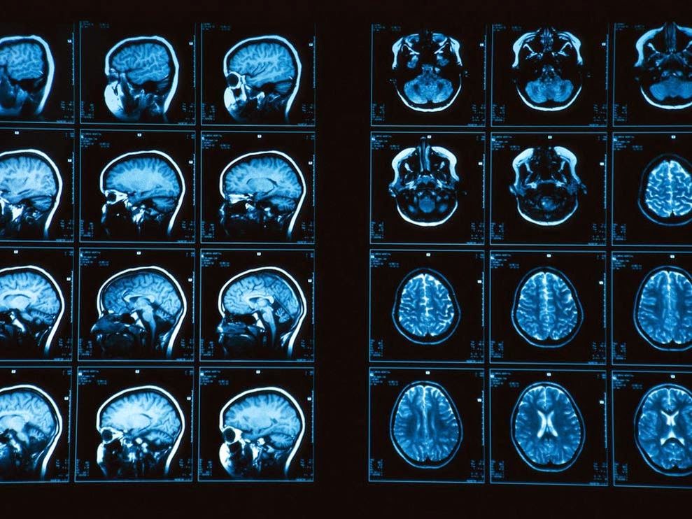
MRI measures how hydrogen nuclei absorb and release energy. Dependent on the location and the environment of the hydrogen atoms the MRI process is able to provide knowledge about the placement of hydrogen atoms in the body and in turn knowledge about the anatomy.


Now for the fun part (see the rules here), using Atom in a sentence by the end of the day:

Serious: Hey Bob, did you know that the atom is the smallest unit that defines the chemical elements and their isotopes?

Less serious: I thought that splitting atoms would produce a large explosion but when I tried using my mom’s perfume “atomizer” it just produced a fine spray and nice smell…


Ok that was a little intense for a Sunday. Watch and listen to Symphony of Science (very cool BBC production) to decompress and I’ll see you in the blogosphere…


Pascal Tyrrell

What Cluster Are You From?

This week and I had the pleasure of presenting to the Division of Rheumatology Research Rounds – University of Toronto. They were a fantastic audience who asked questions and appeared to be very engaged. Shout out to the Rheumatology gang!


So, I was asked to talk about a statistical methodology called Cluster Analysis. I thought I would start a short series on the topic for you guys. Don’t worry I will keep the stats to a minimum as I always do!


Complex information can always be best recognized as patterns. The first picture below on the left certainly helps you realize that it is not a simple task to know someone at a glance.


Now, I guess it doesn’t help that many of you have never met me either! However, you can appreciate that things get a little easier when the same portrait is presented in the usual manner – upright! 


This is an interesting example where the information is identical, however, our ability to intuitively recognize a pattern (me!) appears to be restricted to situations that we are familiar with.


This intuition often fails miserably when abstract magnitudes (numbers!) are involved. I am certain most of us can relate to that. 


The good news is that with the advent of crazy powerful personal computers we can benefit from complex and resource intensive mathematical procedures to help us make sense of large scary looking data sets.


So, when would you use this kind of methodology you ask? I’ll tell you…


1 – Detection of subgroups/ clusters of entities (ie: items, subjects, users…) within your data set.


2 – Discovery of useful, possibly unexpected, patterns in data.


OK, time for some homework. Try to think of times when you could apply this kind of analysis. 


I’ll start you off with an example that you can relate to. Every time you go to YouTube and search for your favorite movie trailer you get a long list of other items on the right that YouTube thinks may be of interest to you. How do you think they do that? By taking into account things like keywords, popularity, and user browser history (and many, many more variables) and using cluster analysis of course! You and your interests belong to a cluster. Cool!


In this series, we will delve into this fun world of working with patterns in data. 


Now that you have peace of mind, listen to The Grapes of Wrath


See you in the blogosphere,


Pascal Tyrrell


MiWord of the Day Is… Cuckoo!

One of my favorite more serious films is One Flew Over the Cuckoo’s Nest. What does Jack Nicholson’s portrayal of a bad guy hoping for easy served time in a mental institution have to do with medical imaging? Well it all starts with the lobotomy. Not to spoil the story, suffice it to say that the movie broaches the topic of lobotomies and how ridiculous they were. Lobotomy was a form of neurosurgery that involved damaging the prefrontal cortex in order to “calm” certain mentally ill patients. Needless to say the procedure was controversial from the beginning (1935 to the early 1970’s) but the author of the discovery, Egas Moniz, was awarded the Nobel Prize in 1949. Maybe not the most sound of decisions by the committee. However, for the time, it was considered progress in a very challenging area of medicine – mental illness. 


OK, medical imaging? Well as it turns out Moniz (do not confuse with St-Moriz, ahhh skiing…) is also known for developing cerebral angiography – a technique allowing the visualization of blood vessels in and around the brain. 


Moniz was interested in finding a non-toxic substance that would be eliminated from the body, but would not be diluted by the flow of blood before the x-ray could be taken. Another requirement is that the substance could not cause an emboli or clot as this would be a bad thing. Moniz played with salts of iodine and bromine and settled on iodine because of its greater radiographic density. And voila, birth of iodinated radiocontrast agents still in use today. Cool.


Supposedly it took him 9 patients to perfect his angiogram technique. Don’t ask about the first 8…

Moral of the story is: lobotomy bad and cerebral angiography good.


Now for the fun part (see the rules here), using Cuckoo in a sentence by the end of the day:

 
Serious: Hey Bob, when I was visiting my aunt in Australia I spied a little bronze cuckoo in her backyard! This could be my “big year“…


Less serious: Someone won a Nobel Prize for developing the lobotomy? Are you cuckoo?
 
 
Listen to Los Lobos (not short for lobotomy but “the wolves” in Spanish) singing La Bamba to decompress and…
 
… I’ll see you in the blogosphere,
 
 
Pascal Tyrrell

Another successful diagnosis thanks to the x-ray…

For more info see Free Birds

Happy Canadian Thanksgiving!!!

A traditional holiday – originating from the native peoples of the Americas – to celebrate the completion and bounty of the harvest. Well, no harvest for me but I will take the time to appreciate some of the successes of our MiVIP program and this blog over the long weekend.


Thanks for being a part of it!


See you in the blogosphere,

Pascal Tyrrell

MiWord of the Day Is… Fluoroscope!

It is hard to believe that the fluoroscope (essentially an x-ray machine used to produce real-time moving images viewed on a screen of the internal structures of a patient) was used to “help” better fit shoes to your feet! From the 1920 to about 1970 you were able to irradiate your feet with x-rays in order to see if you had enough “wiggle-room” in your new shoes! Crazy. 
 
So, the whole concept of Fluroscopy dates back to you know who, Wilhelm Röntgen. We chatted about him here in our blog. He is also responsible for discovering the interesting phenomenon of barium salts fluorescing when exposed to x-rays (see here in our blog). 
 
Basic function of a fluoroscope
Soon after Rontgen’s discovery was announced, Thomas Edison (the light bulb guy) decided he could improve on this whole x-ray thing as these rays were produced by a “glass tube apparatus” – something he knew a lot about. After setting his team to work – he had a team as he was a very successful man in those days following his 1879 patent of the light bulb – they soon discovered the risks of working with x-rays. Edison decided to remove himself (literally!) from x-ray research. But before he did he developed one of the first (and arguably the most advanced in it’s time ) fluoroscopes along with a full line of x-ray kits. He also coined the term “Fluoroscope”. Interesting man…
Fluoroscopes have come a long way over the years and are still used today in areas such as orthopedic surgery, gastrointestinal investigations, and angiography but, of course, the dose of x-rays a patient receives is minimized and closely monitored. Have a look at this machine from Siemen’s. “Beam me up Scotty!”. 
So how did all of these machines suddenly flood the shoe retail industry? Good question. As it happens, following the development of the high vacuum, hot cathode, tungsten-target x-ray tube by William Coolidge in 1913 the interest for a portable and reliable machine increased dramatically with the advent of the First World War. The successful deployment of numerous machines during the war to aid army physicians spurred the manufacturing industry to mass produce them. After the war, the impact the fluoroscope had on army medicine flowed into community practice. 
 
Due to the enormous supply of portable x-ray machines at the time following the end of the war, Dr Jacob Lowe introduced the idea of using a modified portable x-ray machine in the shoe retail industry. Voila, fried feet fricassee for the next 50 years!

Now If were to be interested in using a fluoroscope to look at my feet I may be inclined to use a suit like this gentleman below is sporting…

WW I x-ray protection suit

Now for the fun part, using Fluoroscope in a sentence by the end of the day:


Serious: Bob, did you know that the foot-o-scope was a modified fluoroscope used to view ones feet when fitting new shoes which delivered on average 13 Roentgens for every 20 second exposure?


Less serious: I heard grampa grumbling he can never find shoes that fit right anymore since they banned fluoroscopes in shoe stores. What is a fluoroscope mommy?


Listen to High Heels to decompress and I’ll see you in the blogosphere.


Pascal Tyrrell

MiWord of the Day Is… Radio!

Easy one today! I thought I would give everyone a break as you have all been working very hard on the MiWord of the day in the past weeks. 


So, what does radio have to do with medical imaging? What a great question! The origin of the root word “Radio” is radiant energy. The radio you immediately think of is the one that is attached to your ear most of the time and has a DJ who selects music to play for your entertainment – along with ads to pay for the station’s bills! The use of “radio” to describe this form of wireless communication comes from the word radiotelegraphy


How about if we were simply interested in a medical picture produced by radiant energy? Well you would end up with a radiograph AKA an x-ray! We talked about that word here. Do you see the trend? How about a picture produced by radiant energy in the visible light range of the electromagnetic spectrum? A photograph. Cool.


OK now suppose you are an MD working in the emergency department and someone presents with a lung disorder. What do you do? Generally, you order a chest radiograph. As you zap your patient with x-rays you expect that most of them will pass through the chest area – that is mostly filled with air – unchecked and will proceed to expose the film (or trigger the detector) resulting in a dark area. However, if the lungs become filled with abnormal substances more of the x-rays are blocked and result in a lighter (whiter) radiograph. What would you be looking for?


1- Pus – a combination of bacteria and white blood cells as seen with pneumonia.
2- Edema – fluid that leaks into the lungs as seen with heart failure.
3- Hemorrhage – bleeding into the lung cavity as seen with trauma.
4- a solid mass – as seen in lung cancer.


Today, we have to use “Radio” in a sentence (see rules here). Easy! Here are two examples to help you along:


Serious: Bob, you will need to remove your radio from your person before entering the MRI. No metal objects are permissible in the room.


Less serious: I went for a radiograph today and all they did was have me stand in a room by myself and that was it! What a relief. I thought for a moment I was scheduled for a radio-graft…!


Have a listen to my favorite Radiohead to decompress and…


… I’ll see you in the blogosphere,


Pascal Tyrrell

Face Validity: Who’s Face Is It Anyway?

Yes, I was a big fan of the A-Team. Who wasn’t? Mr. T (I guess that makes me Prof. T…) was always entertaining to watch. Lieutenant Templeton Arthur Peck was suave, smooth-talking, and hugely successful with women. Peck served as the team’s con man and scrounger, able to get his hands on just about anything they needed. Need a refresher? Have a peek here.


Well in a past post 2 Legit 2 Quit we talked about why we assess validity – because we want to know the nature of what is being measured and the relationship of that measure to its scientific aim or purpose. So what if we are uncertain that our measure (a scale for example) looks reasonable? We would consider face validity and content validity. Essentially, face validity assess whether or not the instrument we are using to measure appears to be assessing the desired qualities or attributes based on “the face of it”. Content validity – that was touched on in the previous post – is closely related and considers whether the instrument samples all of the relevant or important content or interest.


So, why the importance of face validity? Whenever you need to interact successfully with study participants there is often a need to:


– increase motivation and cooperation from participants for better responses.
– attract as many potential candidates.
– reduce dissatisfaction among users.
– make your results more generalizable and appealing to stake holders.


These are especially important points to consider when planning a study that involves human subjects as respondents or there exists any level of subjectivity in how data is collected for the variables of interest in your study. 


However, you want to avoid a “Con Man” situation in your study where respondents’ answers are not what they appear to be. As a researcher you need to be aware that there may be situations where Face Validity may not be achievable. Let’s say for instance you are interested in discovering all factors related to bullying in high school. If you were to ask the question ‘ have you ever bullied a classmate into given you his/her lunch money?’ you may have Face Validity but you may not get an honest response! In this case, you may consider a question that does not have face validity but will elicit the wanted answer. Ultimately, the decision on whether or not to have face validity – where the meaning and relevance are self-evident – depends on the nature and purpose of the instrument. Prepare to be flexible in your methodology!


Remember that face validity pertains to how your study participants perceive your test. They should be the yard stick by which you assess whether you have face validity or not.


Listen to Ed Sheeran – The A Team to decompress and…


… I’ll see you in the blogosphere.


Pascal Tyrrell

MiWord of the Day Is Something to “Bragg” About…


OK who hasn’t made rock candy as a kid? No? Give it a try. Maybe you have a little brother or sister you can impress. All you need is a super saturated solution of sugar, a surface for crystal nucleation (string), and lots of time…


Now what if you were to apply this technique to obtain crystals of DNA? I don’t suggest that you eat it as a treat but you could possibly try X-ray crystallography.


The challenge is that DNA, unlike proteins, is an exceedingly large molecule which does not lend itself to crystallisation. The result is a highly viscous suspension of spiderweb-like filaments. However, it is this very suspension that the DNA molecules were deduced to be neatly aligned alongside one another by studying the X-ray diffraction patterns. This initial challenge was successfully overcome by Rosalind Franklin. Her hard work then laid the ground work for Watson and Crick to piece together the puzzle of DNA structure (winning the 1962 Nobel Prize along with Wilkins).


Now the x-ray crystallography imaging technique is no pic-nic! It was first described by the Australian father-and-son duo William Henry Bragg and William Lawrence Bragg. Essentially x-rays are projected onto a crystalline solid and when analyzing the diffraction patterns it is possible to determine how its molecular atoms are positioned in relation to one another. This is due to x-rays having very short wave-lengths (see x-rays in the blog) and the mathematical analysis of predictable diffraction from the three dimensional structure of the crystal. It was Lawrence Bragg who developed the equation to describe this diffraction and is now known as Bragg’s Law. He and his father won the Nobel Prize for this work in 1915. 


I used to listen to New Order back in the day and they are still singing (and dancing?) with a more recent release of Crystal by New Order. So, listen to the song while making rock candy and maybe you too will come up with a brilliant idea worthy of a Nobel Prize by the time the crystals are big enough to eat.


Now if you remember the rules:


1- I introduce and discuss a word.
2- You have to use the word in a sentence by the end of the day. No need to use it in the correct context – actually out of context is more fun and elicits a more entertaining response!


Today, we have to use “Bragg” in a sentence. Here are two examples to help you along:

Serious: “Hey Bob, did you know that if you used a saturated sodium salt solution of DNA instead of sugar to produce crystals we could then do some x-ray crystallography for fun and apply Bragg’s law to determine the molecular structure…”
 
Less serious: “Bob, I don’t want to Bragg but my crystals are way bigger than yours…”


See you in the blogosphere,


Pascal Tyrrell

The Adventures of Alana the Newbie Researcher

Timeline of my ROP experience

Stroke. Atherosclerosis. Carotid stenosis. MRI. Endarterectomy. To some people, these words are, well, just words. But for others, these few phrases could change the course of their lives. During my time in the Research Opportunity Program at the Department of Medical Imaging, I was able to learn all about carotid disease and the current status of carotid imaging modalities. As an added bonus, I did quite a bit of research on the impact that stenosis has on patient care and how new and exciting advances in medical imaging could affect it.


Over the course of four months, I went from being a huge research newbie to becoming an experienced research … trainee. I was introduced to the world of journal article writing, starting from doing elementary literary research all the way to drafting and writing a review article, which was completed entirely from the comfort of my beloved computer. With the help of Doctor Eli Lechtman and my ROP partner Helena Lan (if you haven’t seen her post on her experience you should check it out), we were able to put together 2 review articles that complement one another and will hopefully pose as a good foundation for future research.


Now, you must be thinking, “wow, this sounds like an extremely exciting summer; how could it possibly be better?” Well, my dear readers, it gets even better. One of the greatest things about being a part of this department is the community. The people here are all friendly, wonderful, and welcoming. From having an outing at Centre Island to letting us operate an uItrasound machine (thanks Modele!), I really felt like I was a part of “the gang.” What’s more, it was nice to have someone remember my name when I arrived in the morning, especially when I’m used to being my nameless self in the vast sea of students of our first year lectures (Convocation Hall, anyone?).


When I first sat down to write this post, I couldn’t decide what would be the best way to describe my time doing my ROP. And that’s when it hit me: it was an adventure! I may not have explored unknown lands or survived any near-death experiences, but there was not one day that went by where I wasn’t learning something new. Each day was a new experience, an opportunity to understand something I probably haven’t even heard of before. It still surprises me right now, as I’m writing this post, how much I have learned throughout this program, and the fact that I was able to experience something like this. I would have never thought I could write a review article and do the things I’ve done in only my first year of university. All in all, this was a great experience, and I encourage everyone to try out something like this!


Thanks for reading this, y’all! And if you just scrolled to the bottom of this post and thought “tl;dr,” you can still check out my infographic timeline (there’s pretty pictures)! Also I’d like to give a shoutout to my awesome Professor, Dr. Pascal Tyrrell (check out his latest post), as well as all the lovely members of the Medical Imaging department at U of T for making my ROP experience the way it turned out to be.


Remember to make every day an adventure,


Alana Man 🙂

Helena Lan: Summer 2014 ROP

Helena Lan Summer 2014 ROP

What is research like? If you had asked me this
question several months ago, I would have answered, “You wear a lab coat and
goggles while mixing chemicals or observing organisms. Hopefully something
interesting will happen, so that you get to publish your findings!” Well, after
participating in the Research Opportunity Program (ROP) at the University of
Toronto, I discovered that medical imaging research is more than just
pipetting, and is all the more exciting!

 
So what kind of research is conducted in the medical imaging world? For my ROP, the objective of my project was to evaluate the roles of the non-invasive imaging modalities for diagnosing carotid stenosis. Hence, I engaged in online literature research of the various imaging techniques for assessing this disease. In this process, I also learned to use Zotero to manage all my references, which provides an easy way to generate a bibliography (when the software doesn’t crash every time you open it). After gathering all the pertinent information, I then put together a review article suggesting how a change in the current imaging approach could potentially improve clinical outcome. Who knew a report could be compiled without doing the lab grunt work?     
 
Wait, so this is all a radiologist does? Sitting in front of a computer and typing all day? Of course not! During our time at Sunnybrook Hospital, we got the chance to chat with a radiologist and discovered that she could decide whether patients should be released after taking a look at their diagnostic images. Pretty powerful, eh? That’s not all. We also found out how radiologists identified any abnormalities in patients, as we had the opportunity to work with the VesselMass software which allowed for the delineation of the lumen and vessel wall of arteries on MRI images. Oh, and did I mention we observed an MRI and an ultrasound examination of the carotid arteries, and even got to perform an ultrasound scan ourselves. Super cool!
 
Still craving for more of my ROP experience? Check out my timeline infographic! You will find all the things I learned and all the fun I had there. Last but not least, I’d like to shout out a big THANK YOU to Prof. Pascal Tyrrell and Dr. Eli Lechtman, who guided us every step of the
way. Also, I’m very grateful to Dr. Alan Moody for including us in his research program at Sunnybrook, as well as other members of the VBIRG group who gave us the chance to
participate in various activities. My summer would not have been this fun and meaningful without all of your help!
 
Have fun researching,


Helena Lan